By Tom Watson, clinical analyst with MD Buyline
There is no doubt that new hardware and software solutions unveiled at the 2017 RSNA will have a growing impact on the future of imaging and radiology. But a somewhat different and intriguing perspective was advanced by Dr. Elias Zerhouni, a keynote presenter at the opening of the conference.
Zerhouni is president of Global Research and Development, a member of the executive committee at Sanofi, and the former director of the National Institutes for Health (NIH). His presentation, “Imaging Innovation in 21st Century Biomedicine — Challenges and Opportunities,” posed the intriguing proposition that “What will be is already here.”


Ad Statistics
Times Displayed: 30947
Times Visited: 803 Stay up to date with the latest training to fix, troubleshoot, and maintain your critical care devices. GE HealthCare offers multiple training formats to empower teams and expand knowledge, saving you time and money
This statement seems to suggest that the future is already here, and what we will achieve in the coming years is more about how we integrate and utilize information that we can already acquire than it is about developing totally new data and technology. But I do not believe Zerhouni is suggesting there will be no further innovation in technology or advancement in evolutionary – if not revolutionary – imaging.
“The speed at which things will happen tends to be overestimated,” Zerhouni continued. “We have to participate, not just peripherally, but in the core scientific challenges of today, which are summarized by the tension between the complexity of biological systems and the precision medicine needed in the individual patient population. Imaging innovation is, by its very nature, interdisciplinary.”
While there are many horizons yet to be explored in biomarkers and related areas, the principle that resonated with me was the first, “What will be is already here.” I believe a “works-in-progress” on display at the Philips Healthcare exhibit is a perfect example of that principle being developed in practice.
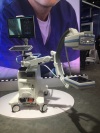
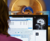
Dr. Atul Gupta, Philips’ chief medical officer as well as an interventional and diagnostic radiologist practicing in Philadelphia, is a key developer and researcher working with Philips on an Augmented Reality (AR) solution. This offers a potential improvement in the workflow and logistics of accessing and integrating the multiple aspects of patient imaging, such as biological results, 3-D image modeling, image fusion of MR, CT and/or ultrasound with interventional x-ray, and even the ability to control the angiography system with a “floating” virtual control board. This latter innovation was available for RSNA attendees to “test fly”.
The combination of Image-Guided Therapy with Augmented Reality (AR) transports the test “pilot” into a data-centric and patient-centric world. With the AR headset on, and in the midst of a virtual patient procedure, the physician could, by using hand gestures, finger movements, and body position, bring up and see virtual representations of CT images, MR images, IVUS, lab results, ultrasound imaging, and hemodynamic monitoring parameters, all in a virtual floating cockpit within his field of view. The result was access to all or many of these parameters without having to turn away from the patient or move to see hardware monitors or other traditional display technologies.