por
John W. Mitchell, Senior Correspondent | December 13, 2017

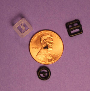
Size comparison between 3-D printed
prosthesis implant and a penny.
Courtesy: RSNA
A team of doctors at RSNA has illustrated the promise 3-D printing may hold for producing exact replicas of the delicate middle ear and curing hearing loss.
By converting 3-D CT images to 3-D printed prosthetics, surgeons were able to correctly match four different-sized implants in different human cadaver ears.
“If we can presume that a highly probable cause of outcome failure with existing prosthesis is due to improper fit, then the ability to create a customized prosthesis that can accurately fit the air/bone gap is less likely to fail,” study author Dr. Jeffrey D. Hirsch, assistant professor of radiology at the University of Maryland School of Medicine in Baltimore told HCB News. “Our study shows that even at a submillimeter level there are minute changes from ear-to-ear that can be accurately represented with 3-D modeling, and are also detectable by otologic surgeons.”


Ad Statistics
Times Displayed: 68650
Times Visited: 2236 Ampronix, a Top Master Distributor for Sony Medical, provides Sales, Service & Exchanges for Sony Surgical Displays, Printers, & More. Rely on Us for Expert Support Tailored to Your Needs. Email info@ampronix.com or Call 949-273-8000 for Premier Pricing.
The technique has the potential to improve a surgical procedure that often fails because of incorrectly sized prosthetic implants, according to Hirsch. In the study, four surgeons performed insertion of each prosthesis into the different middle ears. All four surgeons were able to correctly match the prosthesis model to its intended temporal bone containing the middle and inner parts of the ear.
According to Hirsch, the chances of this occurring randomly are one in 1,296.
“While 3-D printing has been around in the industry for decades, it has only recently [been of] interest in the medical community,” he said. “Radiology is a great avenue for 3-D printing in medicine. As radiologists, we live in a 3-D world, but until recently have only been able to relate to others within the limitations of 2-D representation (flat screen monitors).”
Hirsch explained that 3-D printing brings what radiologists see into the realm of the ordering providers. Being able to see complex anatomic relationships provided by 3-D modeling allows for a new level of learning, comprehension, and medical planning.
The next step for the researchers is to develop a biocompatible material that can be approved for use in live patients. Hirsch said that currently only titanium is regularly used as a prosthetic device in humans. The team, he said, is investigating the use of stem-cell growth using a 3-D printed prosthesis as a platform.
“This could lead to a more permanent solution for ossicular reconstruction (to remedy hearing loss),” Hirsch said. “We propose a procedure to reconstruct a patient’s air/bone gap with a prosthesis that will incorporate into the adjacent bone tissue and create a permanent bone graft bridging the gap.”
Back to HCB News

