por
John R. Fischer, Senior Reporter | May 09, 2018

A new MR detector shows
movement of tendons and ligaments
clearly for the first time
Need a hand?
Researchers at NYU Langone School of Medicine have developed a new, wearable MR component to help patients with hand-related injuries return to these and other activities more quickly by producing, for the first time, clear images of joint movement for enhanced diagnosis.
“CT is really good at visualizing the bones, but it is difficult to see any of the other structures. Video fluoroscopy, generally only provides a 2D projection with very poor soft tissue contrast,” Martijn A Cloos, an assistant professor in the department of radiology at NYU Langone, told HCB News. “MR, on the other hand, provides exquisite soft tissue contrast that can be used to visualize muscle structures, cartilage and nerves.”


Ad Statistics
Times Displayed: 16169
Times Visited: 33 Final days to save an extra 10% on Imaging, Ultrasound, and Biomed parts web prices.* Unlimited use now through September 30 with code AANIV10 (*certain restrictions apply)
But imaging complex, moving joints, such as tendons and ligaments, has been challenging for MR due to its conversion of radio waves by radiofrequency coils into a detectable electric current, with the radio waves producing minute currents, as a result, in the receiver coils. This, in turn, causes the receiver coils to create their own magnetic fields that prevent nearby coils from capturing clean signals to produce such images.
The MR element overrides this limitation using high-impedance coils stitched into a cotton glove.
Unlike conventional coils, designed as low-impedance structures, the new coils block the MR signal from developing a current, thereby preventing the creation of magnetic fields and the interference they cause for neighboring receivers.
The technology consists of high-impedance coils stitched into a cotton glove
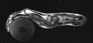
Researchers used the system to examine a hand playing the piano and grabbing objects, finding that it produced clear images of freely moving muscles as well as tendons and ligaments, both of which have historically been difficult to image independently due to their dense protein compositions, appearing as black bands running alongside the bone.
When assessing the hand as it flexed its fingers, the system showed movement of the bands in relation to that of the bones, a finding that researchers claim could help in cataloguing differences in injuries.
The authors hope that the technology will help to construct a more versatile atlas of hand anatomy, produce images of hands in more realistic positions for better surgical guidance, and enhance prosthetic design.
They also suggest that the flexibility of the coils and their immunity to coupling effects make them suitable as comfortable and adaptive MR coils in other applications.
“One particular example application would be a ‘beanie-like' head coil for pediatric patients. We imagine that such a coil may be more comfortable and less intimidating,” Cloos said. “Moreover, such a coil could adapt to the shape and size of the individuals head, thus providing optimal coverage and image quality.”
The findings are compiled in a study published in
Nature Biomedical Engineering.

