por
Lauren Dubinsky, Senior Reporter | April 16, 2018

Siemens Healthineers ACUSON
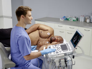
Bonsai cardiac ultrasound system.
From the April 2018 issue of HealthCare Business News magazine
Improvements in imaging technology are not only allowing cardiologists to evaluate patients in ways they never could before, through automation these new tools are simplifying the exam process and saving hospitals valuable time.
Guidelines from the American Society of Echocardiography call for 3-D chamber quantification on all patients undergoing an echocardiogram, but cardiologists have sometimes been known to overlook this time-consuming process when speed and efficiency are top priorities.
Conventional quantification involves the physician hand tracing to evaluate ventricular function, which typically takes about three minutes. There are also limitations associated with that method, such as foreshortening and not being able to visualize the entire endocardial border.


Ad Statistics
Times Displayed: 112448
Times Visited: 6718 MIT labs, experts in Multi-Vendor component level repair of: MRI Coils, RF amplifiers, Gradient Amplifiers Contrast Media Injectors. System repairs, sub-assembly repairs, component level repairs, refurbish/calibrate. info@mitlabsusa.com/+1 (305) 470-8013
This is a prime example of an area where automated tools can simplify the imaging process and enable reproducible results. Meanwhile, the burden on the provider is significantly diminished.
“When you use automated intelligence, once you have acquired the data it doesn’t matter if a physician of two years or a physician with 20 years of experience presses the button because it will give you the same result,” says Dr. Roberto Lang, professor of medicine and director of the cardiac noninvasive imaging lab at the University of Chicago Medicine.
He uses Philips’ Anatomical Intelligence HeartModel tool, which is integrated into his EPIQ cardiac ultrasound systems. It performs 3-D cardiac chamber quantification by simultaneously computing the left ventricle and left atrium volumes from a single volume loop.
“When you have a volume of data, you have more images so you can see more pathology,” says Cassandra Gipson, clinical analyst at MD Buyline. “When you just have 2-D images, it’s limited because it’s a simple still image. With 3-D, you can turn the images around in any direction and see how deep the pathology is.”
Automated cardiac ultrasound imaging also improves workflow in the cardiac catheterization lab because it allows the technologist to have more time with the patient instead of having to stop and reformat.
Lang and his colleagues compared quantification between conventional 2-D exams and HeartModel and found that it reduced the time to obtain results by 82 percent, so if a cath lab typically treats 50 patients per day, he believes they could save over an hour with automation tools.
From a market standpoint, Philips is the leader in the cardiac ultrasound automation tool market and GE Healthcare follows closely behind, according to MD Buyline. Samsung Healthcare, Canon Medical Systems Corporation and Hitachi Healthcare Americas also offer this software on their premium cardiac ultrasound systems.

