por
John W. Mitchell, Senior Correspondent | January 23, 2018
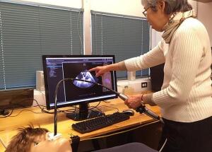
A research cardiologist at the University of Washington School of Medicine (UWSM) has developed the first diagnostic probe ever to allow real-time images to be used to train physicians.
The probe – which looks like a high-tech fishing rod – was developed to provide pre-education to cardiology fellows before they go “live” with patients in an echocardiogram lab.
“A colleague asked me to develop a transesophageal echocardiography (TEE) simulator to prepare the cardiology fellows so that they could better take advantage of the advanced training that the faculty and expert sonographers provide in the echo lab in the cardiology clinic," Dr. Florence Sheehan, director of UWSM Cardiovascular and Training Program, told HCB News. “In other words, she wanted the basic skills to be mastered on the simulator so that the finer points of TEE could be taught while scanning live patients.”


Ad Statistics
Times Displayed: 60606
Times Visited: 1928 Ampronix, a Top Master Distributor for Sony Medical, provides Sales, Service & Exchanges for Sony Surgical Displays, Printers, & More. Rely on Us for Expert Support Tailored to Your Needs. Email info@ampronix.com or Call 949-273-8000 for Premier Pricing.
According to Sheehan, because patients must be sedated for the test, minimizing anesthesia with TEE training improves safety. She also noted that TEE also helps to avoid unproductive sonographer staffing time as training time could be learned on a mannequin instead. And ultimately, the future cardiologists get better training to take better care of patients.
“Our skill metrics give trainees feedback on how they are doing, so they know what progress they are making and also where they need to obtain additional practice. Feedback is the single most important attribute of effective training, reinforces learning, and even enhances retention,” she said.
The UWSM TEE probe is an improvement over current such devices in the market, in that it uses real heart images loaded into a program and acquired by manipulating the rod-like TEE device. Other devices use artificial artist renderings – “cartoons”, according to Sheehan, which she said are not nearly as accurate.
“Since ultrasound is an imaging modality that is quite susceptible to artifact, it is important for trainees to see these artifacts and learn what to do to avoid making a misinterpretation,” said Sheehan. “Our images show what images look like when scanning real patients.”
She noted that the program’s fellowship director decided that the training modules that were available were not accurate enough to use in their program, which motivated Sheehan and her team to develop the UWSM TEE. While the images of the left atrial on their TEE mannequin look like a black and white movie on a television with poor reception, the training is valuable. It provides clues to cardiology residents about a patient’s condition, severity and treatment options. The training module has eight patient cases loaded into the program.
The TEE development began with a grant from The American Heart Association.


Wayne Moore
one of a kind TTE probe
January 24, 2018 09:26
This is not a transthoracic (TTE) probe, it is a transesophageal (TEE) probe.
to rate and post a comment
Gus Iversen
re: one of a kind TTE probe
January 25, 2018 12:18
Good catch, Wayne. We checked with Dr. Sheehan and have updated the article to correct the error. Thanks.
to rate and post a comment