por
John R. Fischer, Senior Reporter | October 24, 2017

Mayo Clinic will be the first practice in
North America to use a 7-Tesla
MR scanner
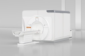
The first clinical 7-Tesla MR imager in North America is predicted to be up, running and scanning patients by the end of 2017 at Mayo Clinic.
The Minnesota-based health system is currently in the process of installing Siemens Healthineers’ MAGNETOM Terra, following
FDA approval for the device earlier this month, making it the first medical center to offer advanced diagnostic imaging through a scanner with the strongest magnetic force available for clinical use.
“I think patients who will benefit the most will be patients that have things that are difficult to assess on a standard MR,” Dr. Kimberly Amrami, chair of the division of musculoskeletal radiology at Mayo Clinic, told HCB News. “We’re going to be using 7-Tesla for those things that require an even higher resolution. The signal-to-noise ratio is directly correlated to the field strength. When we have more signal, we can image smaller structures and still have good images. If the signal to noise is lower and we try to get very, very fine on small structures, it’s difficult, because the images get very noisy.”


Ad Statistics
Times Displayed: 16169
Times Visited: 33 Final days to save an extra 10% on Imaging, Ultrasound, and Biomed parts web prices.* Unlimited use now through September 30 with code AANIV10 (*certain restrictions apply)
The scanner provides more than two times the magnetic field strength of a conventional 3-Tesla scanner to administer ultrafine image resolution of the head and extremities. It is approved for imaging the head and knees and is designed for patients weighing more than 66 pounds.
Mayo radiologists plan to use MAGNETOM Terra to enhance resolution for imaging small lesions in trauma-related micro-hemorrhage patients and for multiple sclerosis lesions that cannot be detected at lower field strength. They will also use it to visualize anatomic sources of seizures in previously undiagnosed patients, and to improve anatomic detail and enhance confidence in noninvasive diagnosis when imaging cartilage and other tissues of the knee, including cartilage implants.
MAGNETOM Terra also provides better visualization of nerves and improves visualization of functional MR imaging by amplifying signals compared with lower field strengths, providing physicians with a better understanding of the location of brain activity in relation to tumors for people undergoing brain surgery.
“I’m really excited about the opportunity to bring this unique tool to our patients; the opportunity to be able to see things we haven’t been able to see or haven’t been able to see that well in the past,” Amrami said. “There are just a lot of tremendously exciting things that will hopefully come out of this. For instance, if we can see the seizure focus better, that hopefully translates into treating that and hopefully curing those problems if possible, or at least getting a definitive answer.”
Back to HCB News

