por
Lauren Dubinsky, Senior Reporter | October 19, 2017

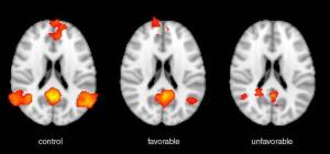
fMR activation
(shown as red-orange)
Advanced MR imaging techniques can help predict neurological outcomes for survivors of cardiac arrest.
A study recently published in
Radiology explains that although current methods are limited, MR imaging can offer more accurate information on long-term recovery potential.
"MR provides high spatial resolution, is multi-parametric, and it lends itself to quantitative analysis," Dr. Robert D. Stevens, lead author and professor at Johns Hopkins University School of Medicine, told HCB News. "The spatial resolution of MR is higher than any other imaging modality."


Ad Statistics
Times Displayed: 107306
Times Visited: 6555 MIT labs, experts in Multi-Vendor component level repair of: MRI Coils, RF amplifiers, Gradient Amplifiers Contrast Media Injectors. System repairs, sub-assembly repairs, component level repairs, refurbish/calibrate. info@mitlabsusa.com/+1 (305) 470-8013
In 2016, there were 559,000 cases of cardiac arrest in the U.S., according to the American Heart Association. Many of the patients who survive are left with severe neurological disabilities that they may or may not recover from.
For the study, Stevens and his team performed diffusion tensor imaging and fMRI exams on 46 patients who were in a coma after suffering cardiac arrest. The exams were conducted within two weeks of the episodes.
They specifically focused on neural connections in the brain such as the default mode network, which is active when a person is not engaged in a task, and a collection of brain regions that select the stimuli that deserve our attention called the salience network.
A year after the cardiac arrest episodes, the team evaluated the patients using the Cerebral Performance Category Scale and found that 11 patients experienced neurological improvements. Functional connectivity was stronger in patients who reached higher levels of independence after one year compared to those who remained significantly dependent.
The team concluded that changes in functional connectivity between brain networks are more accurate predictors of outcomes than structural measures. They also believe that fMRI will be able to help scientists develop therapies and track response to treatment.
The current tools for predicting outcomes include features of the neurological examination, neurophysiology (electroencephalogram, somatosensory-evoked potentials), neuroimaging (CT, MR), and serum biomarkers (for example, neuron-specific enolase).
"Following the introduction of temperature management for patients who suffered cardiac arrest-related anoxic brain injury, it rapidly became apparent that simple prognostic approaches were inaccurate and could lead to misalignment of resources and outcomes," said Stevens.
The researchers don’t expect this MR imaging method to become the standard for predicting long-term neurological outcomes, but they do believe it can give clinicians more confidence when communicating with the families of cardiac arrest patients.
Stevens added that guidelines from multiple societies such as AHA, AAN and ESICM, call for a stepwise algorithm that integrates more than one of the different modalities.

