por
John W. Mitchell, Senior Correspondent | November 10, 2016
qMRI, a modality that extracts quantitative values directly from images — as opposed to qualitative values in normal MR — is just about ready for prime time. After a decade of work, researchers at UC San Francisco's Musculoskeletal Quantitative Imaging Research Interest Group (MQRI) are working to optimize the technique for clinical applications.
"Currently, we are able to identify if patients have regions within their cartilage that are unhealthy - early signs of osteoarthritis including loss of proteoglycan and loosening of the collagen network," Richard Souza, PT, Ph.D., vice chair for research, Department of Radiology and Biomedical Imaging at the University of California, San Francisco told HCB News. "These measures are observable long before the standard clinical X-ray diagnosis strategy would identify disease."
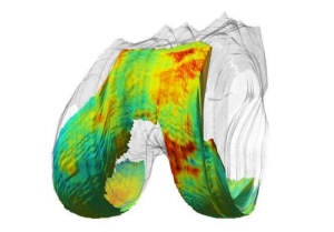
According to Souza, by combining biomechanics testing with qMRI, osteoarthritis risk factors can be identified. These values are translated to a color scale to produce full color, information-rich images, with red indicating areas of degenerating cartilage. This occurs directly from the image with specialized programming, without a radiologist reading.


Ad Statistics
Times Displayed: 16169
Times Visited: 33 Final days to save an extra 10% on Imaging, Ultrasound, and Biomed parts web prices.* Unlimited use now through September 30 with code AANIV10 (*certain restrictions apply)
One in 20 adults suffers from osteoarthritis, caused by joint wear and tear. UCSF is one of only two medical centers in the world where clinicians are at work to study and develop qMRI preventive therapies for osteoarthritis. Souza and his qMRI colleagues, including Sharmila Mujumdar, Ph.D., director at qMRI, have been working on qMRI for more than a decade.
"It's extremely valuable to be able to quantify cartilage composition, because subtle composition changes are reversible," said Souza. "qMRI allows you to look at cartilage in a healthy enough stage when you can still reverse the damage. That opens up a whole world of interventions."
Standard MR provides only the morphology of cartilage, and by the time large lesions are visible, the damage is irreversible.
Early intervention allows clinicians to intervene sooner to help patients. Biochemical changes to cartilage identified by qMRI can be treated through physical therapy and/or surgery. This includes therapy to adjust walking gaits or interventional surgery to re-contour lesions or repair tears to keep injuries from worsening.
"As a physical therapist, the ability to noninvasively evaluate the health of articular (smooth) cartilage is an extremely exciting development," said Souza.
Souza said he, Majumdar and Dr. Alan Zhang, an orthopedic surgeon, have all discovered new insights and options in caring for patients via the qMRI imaging project.
"In addition, qMRI provides a unique opportunity to evaluate response to treatment with a highly sensitive imaging biomarker," said Souza. "Treatment protocols can be evaluated in an accelerated manner using these sensitive new tools".

