por
John W. Mitchell, Senior Correspondent | June 30, 2015
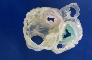
Congenital heart experts have printed the first 3-D heart integrating CT and three-dimensional transesophageal echocardiography (3DTEE) imaging. The team at Spectrum Health Helen DeVos Children’s Hospital in Grand Rapids, Mich. designed the heart as an aide for diagnosing cardiovascular problems and for interventional or surgical planning.
“This first step was testing the feasibility of integrating two or more imaging modalities to print a hybrid 3-D heart model. The next step is to integrate MRI and 3-D ultrasound data for 3-D printing in preparation for surgical planning,” Bennett Samuel, clinical research nurse and one of the study authors told DOTmed News.
He added there is a study in the works to evaluate the efficacy of 3-D printing in surgical and interventional treatment planning. Samuel worked with lead author and cardiac sonographer, Jordan Gosnell, and Dr. Joseph Vettukattil, co-director of the Congenital Heart Center and Division Chief of Pediatric Cardiology at Helen DeVos Children’s Hospital, and senior study author.


Ad Statistics
Times Displayed: 16169
Times Visited: 33 Final days to save an extra 10% on Imaging, Ultrasound, and Biomed parts web prices.* Unlimited use now through September 30 with code AANIV10 (*certain restrictions apply)
The 3-D printed heart was made using the imaging studies of an actual patient with a congenital heart defect. The modeling required the patient to undergo a CT study of 20-30 minutes and a 3-D TEE for about 30 minutes. He said the 3-D model offers immediate possibilities as a teaching tool.
“The 3-D heart model may be used to teach patients and their family members about their congenital heart defect and the plan for repair. Currently, a drawing on a piece of paper or whiteboard is used to explain the procedure to the patient,” Samuel explained.
He also said the 3-D model would be a valuable device to teach medical students, residents, nurses and other medical professionals about their patient’s condition and how to provide the best care.
The Helen DeVos Children’s Hospital team developed the hybrid 3-D heart model in collaboration with the Belgium-based company, Materialise, a global provider of 3-D printing software and technology with 166 industrial grade machines printing more than 2,000 parts a day for clients ranging from aeronautics to consumer products.
At press time, Dr. Vettukattil was attending a meeting to present the 3-D heart research project to 837 cardiologist colleagues from 67 countries at CSI – Catheter Interventions in Congenital, Structural and Valvular Heart Disease in Frankfurt, Germany. Samuel said the development of the hybrid 3-D heart offers many possibilities the team is just considering. But ultimately, patients will benefit.
“There is a possibility for customized patient care as each congenital heart defect differs from patient to patient,” said Samuel.

