por
Diana Bradley, Staff Writer | April 25, 2012

The packaged
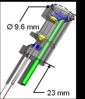
endoscope overlaid with
the optical system.
For the first time, an endoscopic medical device fitted with a femtosecond laser "scalpel" enabling surgeons to spare healthy cells by only removing unwanted tissue has been developed by a research team from the University of Texas at Austin.
"This device has a lot of unique properties and will create the next generation of surgical tools," the project's principal investigator, Dr. Adela Ben-Yakar, told DOTmed News. "This will allow doctors to carry out treatments that were not possible before and may even improve current ones."
With traditional scalpels or surgical lasers, most medical operations end up with the accidental destruction of some healthy tissue - particularly when surgeons are dealing with delicate areas like the brain, throat, and digestive tract.


Ad Statistics
Times Displayed: 32709
Times Visited: 865 Stay up to date with the latest training to fix, troubleshoot, and maintain your critical care devices. GE HealthCare offers multiple training formats to empower teams and expand knowledge, saving you time and money
While tabletop femtosecond lasers have already been in use for the past 10 years in eye surgery, Ben-Yakar's team is working on a way to utilize the technology to repair vocal cords, remove small spinal cord tumors and other bad tissue.
"Currently there is minimal use of lasers with spinal cord surgeries," Ben-Yakar says. "This new tool will cut out spinal cord tumors without causing any carbonization. It's very clean; it just vaporizes whatever the laser touches."
Along with working on a project involving nanosurgery on brain neurons and synapses and cellular structures such as organelles, Ben-Yakar's group is currently developing a new treatment for treating scarred vocal folds with a larynx-tailored probe.
"Anything from smoking to talking too much to sickness and cancer treatments can cause vocal folds to scar, which can lead to voice disorders," says Ben-Yakar. "There is no treatment for this currently and we are trying to develop the only effective treatment."
Coupled with a mini-microscope, the team's device includes a laser capable of generating pulses of light a mere 200 quadrillionths of a second in duration. The specialized microscope uses "two-photon fluorescence" - an imaging technique, which relies on infrared light penetrating up to one millimeter into living tissue. This enables surgeons to target anything from individual cells to even tinier parts like cell nuclei. Thinner than a pencil and less than an inch long, the entire device can fit into large endoscopes; for example: those used for colonoscopies.
Laboratory tests have already taken place with human breast cancer cells, pig vocal cords and rat-tail tendons.
"We are the only group who pursues this kind of technology development; we are leading the field in that sense," says Ben-Yakar. "In two years, we can start pre-clinical studies; maybe even less if everything comes together. Clinical testing should last around five years before we can get FDA approval for human use."
The researchers will present their work -- supported by the National Science Foundation and by the University of Texas Board of Regents Texas Ignition Fund -- at this year's Conference on Lasers and Electro Optics in San Jose, Calif., taking place May 6-11.

