by
John R. Fischer, Senior Reporter | June 26, 2023
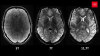
German researchers were able to visualize a proton beam and determine its range during irradiation with real-time MR. (Photo courtesy of Stephan Wiegand/Hochschulmedizin Dresden)
While designed to destroy tumors and preserve healthy tissue, the inability to visualize the beam and map out its range in relation to a patient’s anatomy limits the precision of dose control in proton therapy.
In Germany, researchers at the National Center for Radiation Research in Oncology – OncoRay, operated by the Helmholtz-Zentru Dresden-Rossendorf (HZDR), have observed the proton beam using real-time MR scanning. With more research, they say their technique could potentially make proton therapy precise enough in a few years to treat mobile tumors with higher doses and without damaging healthy tissue.
While irradiating a fluid-filled phantom, the group used an in-beam MR prototype to visualize the beam within the liquid, the first example of real-time MR imaging during proton irradiation, and captured images showing increased depth of penetration as proton energy increased, indicating that MR signal strength also rose.


Ad Statistics
Times Displayed: 174449
Times Visited: 3180 For those who need to move fast and expand clinical capabilities -- and would love new equipment -- the uCT 550 Advance offers a new fully configured 80-slice CT in up to 2 weeks with routine maintenance and parts and Software Upgrades for Life™ included.
“In clinical practice, the proton beam is delivered in water phantoms prior to the actual patient treatment in order to check various beam properties. So far, these QA measurements have been indirect and time-consuming, but with the in-beam MR scanner the proton beam can now be imaged directly during QA measurements,” Aswin Hoffmann, professor and head of the experimental MR-integrated Proton Therapy Group Institute of Radiooncology – OncoRay, told HCB News.
According to Aswin, they need to better understand the MR contrast mechanism behind their technique before applying it in vivo during dose delivery to patients. To do this, they are installing a new large-scale MR system in the OncoRay building to perform proton irradiation and real-time MR simultaneously and directly adjust the direction and strength of the magnetic field relative to the patient.
“We need to investigate whether it is possible to enhance the sensitivity of this method to extend its applicability to tissue materials and clinical dose levels and dose rates,” he said.
They next will investigate if the local MR signal magnitude loss observed in the beam region depends on beam-induced convection, and if signal phase information enhances sensitivity based on signal magnitude. Aswin hopes to use the technique in a few years to monitor patient treatments.
The findings were published in the
Proceedings of the National Academy of Science.