by
John R. Fischer, Senior Reporter | April 03, 2024
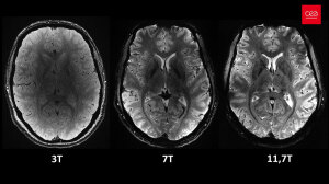
Axial view of the human brain, with the same acquisition time but different magnetic field strengths (Photo courtesy of CEA)
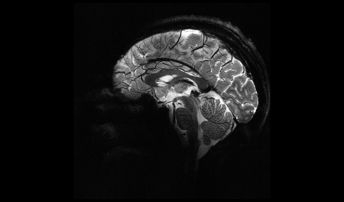
After more than 20 years, the first images of human brains produced with the most powerful MR magnet in the world, the 11.7T Iseult MR machine, have come out, providing ultra-detailed anatomical insights that are expected to shed light on the mechanism of the mind, its neurological states, and the effects that various chemicals and conditions have on it.
Housed at NeuroSpin, a brain imaging research center on the CEA Paris-Saclay site, under Le CEA (the French Alternative Energies and Atomic Energy Commission), the scanner assessed twenty healthy volunteers’ brains, capturing images for each one in about four minutes. This is significantly less time than is currently needed with standard 1.5T and 3T clinical machines used in hospitals. The resolution and contrast that the images were captured with could only be achieved on 1.5T and 3T MR scans conducted over more than two hours, said scientists at Neurospin.
The images have a 0.2 mm in-plane resolution and 1 mm slice thickness, equivalent in volume to a few thousand neurons. The researchers at Le CEA say that it will provide previously unattainable information about different parts of the brain and show how it forms mental representations and the neuronal signatures associated with its state of consciousness. It will also provide a diagnostic understanding of Alzheimer’s, Parkinson’s, and other neurodegenerative diseases.


Ad Statistics
Times Displayed: 175230
Times Visited: 3189 For those who need to move fast and expand clinical capabilities -- and would love new equipment -- the uCT 550 Advance offers a new fully configured 80-slice CT in up to 2 weeks with routine maintenance and parts and Software Upgrades for Life™ included.
“We still need several years of research to develop and improve our acquisition methods and ensure the data has the highest quality possible. Our goal is to investigate neurodegenerative diseases by 2026 to 2030, as well as other diseases that fall more under psychiatry, such as schizophrenia and bipolar disorders,” said Nicolas Boulant, head of the Iseult project and director of research at the CEA, in a statement.
11.7T magnetic field vs. 1.5 and 3 T for conventional MR machines in hospitals. (Photo courtesy of CEA)
Along with better assessing various neurological disorders and conditions, the solution can trace the distribution of chemicals that are hard to capture in images at lower magnetic fields due to their weak signals, showing how they interact with the brain and their efficiency. This includes lithium for bipolar disorder, and glucose and glutamate, molecules associated with metabolism that also provide information about brain diseases such as gliomas and neurodegeneration.
Beginning in the early 2000s as part of a Franco-German partnership, one of the main pillars of the project was to build the most powerful MR scanners in the world. The magnet was designed by Alstom, now part of GE HealthCare, and weighs 132 tons. It has 182 km of superconducting wires, with 1,500 amperes running through its coils. The temperature at which it is kept is -271.35 °C, cooled by 7,500 liters of liquid helium.
Siemens Healthineers installed the additional peripheral equipment for the MR system, and the University of Freiburg in Germany developed new technologies and methods for performing ultrahigh-field MR scans with it. Also involved in its formation was Guerbet, which used the platform at Le CEA to assess and select compounds that demonstrate significant potential for use in humans.