by
John R. Fischer, Senior Reporter | November 23, 2021
GE Healthcare's silicon-based photon counting CT system is being clinically evaluated for use
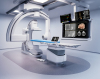
The world’s first silicon-based photon-counting CT system, designed by GE Healthcare, is now under clinical evaluation in a pilot study overseen by Karolinska Institutet and MedTechLabs.
In photon-counting CT, detectors measure each individual X-ray that passes through a patient’s body. This allows it to collect more detailed information to better visualize organ structures, improve tissue characterization, make more accurate material density measurements and lower radiation dose. It is expected to significantly enhance imaging in oncology, cardiology, neurology and for many other clinical CT applications.
GE used pure silicon detectors developed by Prismatic Sensors, which it acquired in November 2020 for this very endeavor. Compared to other photon-counting CT detector materials, silicon is purer, more abundant and has broad manufacturing infrastructure. The main objective of the trial is to study the performance of the machine’s Deep Silicon detector technology, which combined with photon-counting CT has the potential to produce stronger spatial resolution. The study will also compare the performance of the photon-counting CT system with Deep Silicon to standard CT technology and collect insights for optimizing image processing.


Ad Statistics
Times Displayed: 172766
Times Visited: 3129 For those who need to move fast and expand clinical capabilities -- and would love new equipment -- the uCT 550 Advance offers a new fully configured 80-slice CT in up to 2 weeks with routine maintenance and parts and Software Upgrades for Life™ included.
"What makes photon-counting CT with Deep Silicon detectors so unique is that the resolution of the images can be greatly increased, and the dose of radiation reduced, which is particularly important for pediatric patients. You can, for example, determine the degree of constriction in a calcified blood vessel much more accurately, see smaller blood vessels than is currently possible and more easily identify a stroke in certain parts of the brain," Staffan Holmin, professor at Karolinska Institutet, consultant at ME Neuroradiology at Karolinska University Hospital, and clinical evaluation leader responsible for testing and optimizing the technology, told HCB News.
He adds that the system can also be used to image vascular pathologies and see malignant changes at earlier stages when treatment is more effective.
Silicon detectors have traditionally been too thin to stop and collect a sufficient number of X-ray photons when placed in a “face on” position. GE got around this by placing the silicon sensors “edge on”, which allows the detectors to handle the very high photon flux from the CT’s X-ray tubes. The sensors can count hundreds of millions of CT photons per second to create sharper images than standard CT.