by
John R. Fischer, Senior Reporter | January 08, 2020
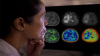
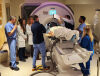
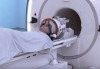
To test the accuracy of the CNN, researchers enrolled 278 patients in a prospective clinical trial to undergo brain tumor resection or epilepsy surgery at three university medical centers. Brain tumor specimens were biopsied from patients, split intraoperatively into sister specimens, and randomly assigned to a control arm or an experimental arm.
Those in the control arm, which represented the current standard practice, underwent specimen processing, slide preparation by technicians, and interpretation by pathologists, a process which takes 20-30 minutes. The experimental arm was performed intraoperatively, with the CNN conducting the entire process from image acquisition and processing to diagnostic prediction. The pathologist-based interpretation was 93.9 percent, compared to the AI-based diagnosis, which scored 94.6 percent accurate.
The unique nature of diagnostic errors in the experimental group compared to those in the control group suggests that a pathologist using the approach could score close to 100 percent accuracy. The system’s strong diagnostic ability could also benefit centers that lack available expert neuropathologists.
"SRH will revolutionize the field of neuropathology by improving decision-making during surgery and providing expert-level assessment in the hospitals where trained neuropathologists are not available," said co-author Dr. Matija Snuderl, an NYU Langone neuropathologist, and molecular pathologist, and associate professor in the department of pathology at NYU Grossman School of Medicine, in a statement.
The findings were published in
Nature Medicine.
Back to HCB News