by
John R. Fischer, Senior Reporter | January 23, 2019
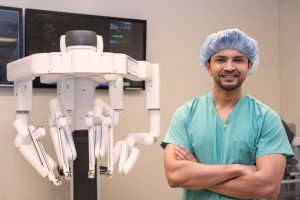
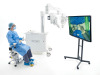
Northwestern Memorial used the da Vinci
Xi Surgical System to perform the first
robotic-assisted lung volume reduction
surgery in the U.S.
Northwestern Memorial Hospital has become the first provider in the U.S. to perform robotic-assisted lung volume reduction surgery, using the da Vinci Xi Surgical System.
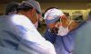
The system was used by hospital staff to precisely target and remove the diseased, emphysematous tissue within the lungs of a patient with severe emphysema, reducing pain, scarring and risk of infection, and providing a shorter recovery time.
"Milestones such as these give us great pride in the relentless pursuit to always provide better surgical treatment options for patients who are in need," Dr. Ankit Bharat, MD, surgical director of the Lung Transplant Program & ECMO at Northwestern Memorial Hospital, told HCB News. "I’m ecstatic to be able to now offer this minimally invasive option to our patients who are seeking a surgical treatment option for COPD. Combining this medical innovation with exemplary patient care opens doors for more patients to become good candidates for robotic lung volume reduction surgery because of the decrease in risk."
Emphysema is a form of chronic obstructive pulmonary disease, a condition that affects 16 million Americans currently, according to the U.S. Centers for Disease Control and Prevention.
Traditional surgical techniques require a larger chest incision to access the lungs, whereas robotic LVRS relies on three eight-millimeter incisions which reduces scarring, risk of infection and potentially, the length of stay within a hospital following the procedure.
The system enables surgeons with specialized training in robotic surgery to oversee procedures from a computer console, looking through a stereoscopic, high-definition monitor to peer inside the patient. This provides a more detailed 3D view of the operating area compared to the human eye.
Comprising the solution is a tower containing four arms. One carries the system’s 3D cameras, while the other three can hold a multitude of surgical equipment, with a computer controlling and replicating each one’s movement to match those of an operating surgeon.
During surgery, the tower is placed directly over the patient. Using master controls, the surgeon directs the arms to make three small incisions on the right side of the chest to access the lungs and remove the diseased cysts. This allows the remaining, healthy lung tissue to support optimal breathing, with the diaphragm, chest wall and rib cage returning to a more normal state.
"Surgical therapies are evolving to enhance precision using minimally invasive platforms," said Bharat. "Incorporating artificial intelligence in the imminent future is likely to enhance our surgical decision-making and better manage intraoperative variability. Additionally, the multidisciplinary approach used in our case, incorporating experts from both pulmonary medicine and thoracic surgery, will be increasingly incorporated to provide the most effective and improved treatment options to our patients."
The system was
adopted just a little over a year ago by the Montreal Heart Institute for use in cardiac surgery.
It is FDA-cleared and CE-marked.