by
John R. Fischer, Senior Reporter | May 14, 2018
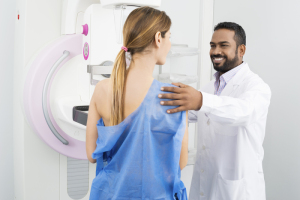
A new study says that automated
breast-density assessments are
just as accurate as those conducted
by radiologists
Automated breast-density evaluation is just as accurate in predicting the risk of breast cancer among women as are examinations conducted by radiologists.
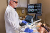
That’s what researchers at the University of California, San Francisco and Mayo Clinic claim in the largest study to compare both methods in assessing patients with screening-detected malignancies and those with interval invasive cancers diagnosed in clinical exams a year after receiving a clean bill of health from a mammogram.
“We found that automated and clinical breast density assessment methods were equally accurate at predicting both the risk of cancer detected through mammography screening and the risk of interval invasive cancer. Automated density measures are more reproducible across radiologists and facilities,” Karla Kerlikowske, a lead author of the study and a UCSF Professor of Medicine, told HCB News. “Using automated measures will allow accurate identification of women who have dense breasts and are at high risk of an interval cancer so these women can have appropriate discussions of whether supplemental imaging is right for them.”
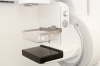
Dense breast tissue can increase tumor aggressiveness and obscure the presence of tumors, preventing mammograms from detecting them and creating a higher risk for women with dense breasts of incurring advanced-stage breast cancer. Those with interval cancer are especially at risk with their condition likely to remain undetected for longer periods of time.
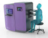
Using Volpara software to measure volumetric breast density on raw digital images, researchers examined 6,369 women matched by age, race, state of residence, screening date and mammography machine from screening practices in San Francisco and Rochester, Minnesota.
Of the participants, 1,609 were diagnosed with cancer within a year of receiving a positive mammogram; 351 had invasive cancer; and 4,409 were in the control group.
The authors found that women with extremely dense breasts who underwent an automated BI-RADS assessment were 5.65 times more likely to be diagnosed with interval cancer and 1.43 times at higher risk of being diagnosed through screening than women with scattered fibroglandular densities, the most common density category for women at average risk.
The results of the study follow a recent number of states that have passed legislation this year, requiring patient-issued mammography reports to specify if a woman has dense breast tissue so that she and her provider may determine if additional screening options are necessary. The most recent
include Utah, Washington, Florida and Wisconsin,
making the total number to do so 35.
Kerlikowske says both methods predicted interval cancer more strongly than screen-detected cancer, illustrating the need to separate the two when assessing risk.
“Automated and clinical BI-RADS density is a strong predictor of interval cancer, much stronger than for screen-detected breast cancer,” she said. “Separate risk models should be developed to predict interval cancer and screen-detected breast cancer and density measures for these models can be up to five years before cancer diagnosis.”
The study was funded by the National Cancer Institute and was published in the Annals of Internal Medicine.