by
Lauren Dubinsky, Senior Reporter | January 16, 2018
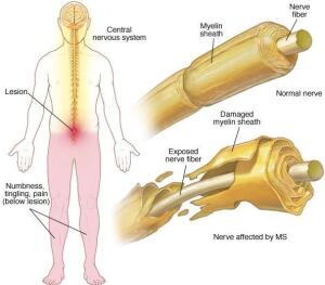
Researchers have developed a PET tracer that can measure damage caused by multiple sclerosis.
“Depending of the nature of a lesion, the treatment choice may be different,” Pedro Brugarolas, first author of a paper published in
Scientific Reports and faculty member at MGH/Harvard Medical School, told HCB News. “This new PET tracer may be able to clarify the nature of the lesion.”
The loss or damage of a cellular sheath that insulates nerves called myelin is a hallmark of MS. Physicians use MR to image demyelination, but it doesn’t provide quantitative data and cannot distinguish between demyelination and inflammation.


Ad Statistics
Times Displayed: 365811
Times Visited: 7093 Quality remanufactured Certified Centrifuges at Great prices! Fully warranted and backed by a company you can trust! Call or click for a free quote today! www.Centrifugestore.com 800-457-7576
Brugarolas and his colleagues at the University of Chicago Medicine and the National Institutes of Health investigated a PET tracer designed to target voltage-gated potassium channels, a protein found on demyelinated axons.
They started with an existing drug to treat MS called 4-aminopyridine, which can bind to exposed potassium channels. In mouse models with MS, they showed the drug accumulates in the demyelinated areas of the central nervous system.
The team then studied several fluorine-containing derivatives of 4-aminopyridine for binding to K+ channels. They uncovered that 3-fluoro-4-aminopyridine (3F4AP) has the ideal properties, so they labeled the molecule with fluorine-18, which is easily detected by PET.
In partnership with scientists at NIH, the research team conducted a study in healthy primates and found that radiolabeled 3F4AP enters their brains and localizes to areas where there is little myelin.
The researchers concluded that the approach can provide complementary information to MR exams. It has the potential to track responses to remyelinating therapies and also help determine the damage that other central nervous system disorders do to the myelin.
“MR has better resolution than PET so it produces finer images,” Brugarolas said. “MR with gadolinium contrast can also inform if there's disruption of the blood-brain barrier so there is information that can be obtained from an MR that cannot easily be obtained from PET. Typically, PET is combined with MR so that you get the best of both worlds.”