by
John R. Fischer, Senior Reporter | December 18, 2017
Researchers have developed a non-
invasive approach using radiotherapy
to halt life-threatening arrhythmias
Patients with life-threatening heart arrhythmias may soon have a noninvasive treatment option in the form of radiotherapy.
Physicians at Washington University School of Medicine in St. Louis have developed a new approach for treating ventricular tachycardia, an irregular heart rhythm condition that puts people at risk for sudden cardiac death, by applying stereotactic radiation directly to the heart. Their findings were compiled in a study published in
The New England Journal of Medicine.
"Catheter ablation is the standard of care. While it can be quite effective in many patients, it has high rates of recurrence - 50 percent in some series - and the potential for death or severe short term toxicity, particularly in the sicker patients," first author Phillip S. Cuculich, an associate professor of medicine and cardiologist, told HCB News. "When patients have a recurrence after catheter ablation, or are too sick for catheter ablation, they have few options. Our totally noninvasive method of mapping the arrhythmia and delivering radiotherapy to the target in under 15 minutes has the chance to significantly improve short term safety for this patient population. We remain committed to monitoring for the long term effects of this treatment.


Ad Statistics
Times Displayed: 369856
Times Visited: 8588 Quality remanufactured Certified Centrifuges at Great prices! Fully warranted and backed by a company you can trust! Call or click for a free quote today! www.Centrifugestore.com 800-457-7576
An estimated 300,000 deaths per year in the U.S. are credited to ventricular tachycardia, the leading cause of sudden cardiac death. Standard therapy consists of medication and invasive procedures that involve threading a catheter through a vein into the heart. While defibrillators can help save the lives of such patients, the shock applied can often be a traumatic experience.
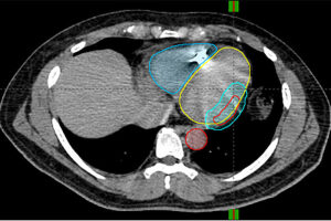
The approach, the first entirely noninvasive process proposed for this condition, consists of imaging the heart using MR, CT or PET scans. Electrocardiographic imaging is then applied in which electrical mapping overlays scar mapping for pinpointing the location of arrhythmias and where they may progress. Radiation is then administered over a period of 10-15 minutes.
Researchers tested their approach on five participants who either were unable to undergo catheter ablation because of other high-risk conditions or did so only for their arrhythmias to return. Together, all five had experienced more than 6,500 episodes of ventricular tachycardia in the three months preceding radiation therapy with the average number per patient being 1,315 in a range of 5-4,312.
Radiation was applied using Varian TrueBeam, a linear accelerator with stereotactic radiation capabilities, administering a single dose on par with what might be provided to an early-stage lung tumor patient. The amount of doses applied ranged between one and five.