by
Thomas Dworetzky, Contributing Reporter | November 02, 2017
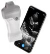
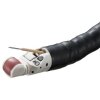
An ultrasound machine with a Nintendo Wii controller chip taped to it just might change the rules of 3-D scanning.
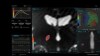
For a fraction of the cost of a conventional 3-D scanning devices, such as a MR and CT machines, the chip is able to locate the ultrasound controller in space so that software can then combine the positioning data and the ultrasound image to create a 3-D composite – similar in quality to a CT or MR scan.
“With 2-D technology you see a visual slice of an organ, but without any context, you can make mistakes,” says
technology co-creator Dr. Joshua Broder, an emergency physician and associate professor of surgery at Duke Health. “These are problems that can be solved with the added orientation and holistic context of 3-D technology. Gaining that ability at an incredibly low cost by taking existing machines and upgrading them seemed like the best solution to us.”
The brainstorm hit the physicians when he was fooling around with his son's Nintendo Wii system.
The game could precisely track the controller's position in space, so why not do the same for the ultrasound?
He experimented for a year and then connected with Matt Morgan, at the time an undergrad at Duke’s Pratt School of Engineering, and biomedical engineering professors Carl Herickhoff and Jeremy Dahl, now at Stanford.
To use the ultrasound machine to create a 3-D image, a handle-like plastic object with the positioning microchip in it is snapped onto the probe.
The data from the probe and microchip are massaged together by software in a laptop to produce a 3-D image.
The system's affordability, ease-of-use and portability could make the device a lifesaver.
“With trauma patients in the emergency department, we face a dilemma,” Broder said. “Do we take them to the operating room not knowing the extent of their internal injuries or bleeding, or do we risk transporting them to a CT scanner, where their condition could worsen due to a delay in care? With our new 3-D technique, we hope to demonstrate that we can determine the source of bleeding, measure the rate of bleeding right at the bedside and determine whether an operation is really needed.”
Another likely patient candidate is a neonate in intensive care. Although smaller scanners for the NICU are being designed, they are rare, expensive and still difficult to use in tight spaces.
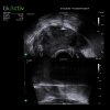
Broder and colleagues have used their device to gather 3-D images of the brain of a baby with hydrocephalus.
The baby slept through the procedure and never noticed it.
“Ultrasound is such a beautiful technology because it’s inexpensive, it’s portable, and it’s completely safe in every patient,” said Broder. “And it’s brought to the bedside and it doesn’t interfere with patient care.”
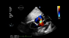
The development team is now working out methods to expand the power of their modified 3-D ultrasound – including a way to image a beating heart.
“In emergency medicine, we use ultrasound to look at every part of the body — from looking at blood vessels that we put catheters into, to checking on a trauma patient to see where they’re bleeding,” Broder said. “In this case we can augment 2-D machines and improve every one of those applications. Instead of looking through a keyhole to understand what’s in the room, we can open a door and see everything in front of us.”