Over 900 Cleansweep Auctions End Tomorrow 05/02 - Bid Now Over 800 Total Lots Up For Auction at Four Locations - TX 05/03, TX 05/06, NJ 05/08, WA 05/09
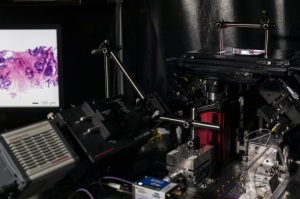
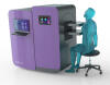
It takes less than 30 minutes for a new light-sheet microscope to scan excised breast tissue for cancer cells in mid-lumpectomy and yield results comparable to traditional pathology approaches that can take days.
This could spare women the need to undergo additional procedures to remove cancerous tissue missed the first time.
“Surgeons are sort of flying blind during these breast-conserving surgeries,” said University of Washington mechanical engineering professor Jonathan Liu in a report of the new device. “Oftentimes they’ve left some tumor behind which they don’t know about until a few days later, when the pathologist finds it.”
At present from 20 to 40 percent of women need additional operations before all cancerous tissue is removed.
“If we can rapidly image the entire surface or margin of the excised tissue during the procedure, we can tell them if they still have tumor left in the body or not. And that would be a huge benefit to cancer patients,” Liu said.
The new microscope is described in a paper published June 26 in Nature Biomedical Engineering.
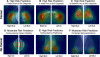
Unlike present processes and microscopic approaches, it doesn't destroy tissue. Instead it creates a “sheet of light to optically "slice" through and image a tissue sample without destroying any of it,” according to a UW statement. Thousands of high-resolution 2-D images are created and then “stitched” together to create a 3-D image of the tissue that can be zoomed in on to view cells in great detail.
“Slide-based pathology is still an analog technique, much like radiology was several decades ago when X-rays were obtained on film. By imaging tissues in 3-D without having to mount thin tissue sections on glass slides, we are trying to transform pathology much like 3-D X-ray CT has transformed radiology,” Liu said. “While it is possible to scan microscope slides for digital pathology, we digitally image the intact tissues and bypass the need to prepare slides, which is simpler, faster and potentially less expensive.”
By contrast, traditional pathology approaches have “changed little over the past century,” noted co-author Nicholas Reder, chief resident and clinical research fellow in UW Medicine’s Department of Pathology. “This light-sheet microscope represents a major advance for pathology and cancer patients, allowing us to examine tissue in minutes rather than days and to view it in three dimensions instead of two — which will ultimately lead to improved clinical care.”