by
Lee Nelson, Contributing Reporter | November 05, 2015
From the November 2015 issue of HealthCare Business News magazine
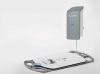
The other solution is onboard image storage capacity. These types of detectors can store from 100 to 400 images. The result is something of a cross between CR and DR: the detector needs to go to a reader where the images are downloaded, but because it’s DR, you get the advantage of low-dose capability.
Reduction of dose and better image quality
The reason many medical facilities are switching to DR, and sometimes from film to CR, is to get better image quality and to reduce radiation dosage given to patients during the procedure. Agfa Healthcare received clearance from the FDA last year to claim its DR systems reduced dose radiation up to 60 percent compared to film. And you will find all the OEMs citing dose reductions of 40 to 60 percent, or more, with DR.
“We have affordable low dose in CR as well as DR,” says
George Curley, senior sales marketing manager at Agfa, “and we have new CR systems that give you better image quality at a lower dose.” However, he admits, customers are moving from CR to DR to gain efficiency, especially in fast-paced medical centers. “It helps managers to justify the initial expense of going to DR if you can get more efficient on 70 percent of all your exams,” Curley says. “That’s huge, because it is the biggest volume of procedures.”
Dose reduction is a double-edged sword, according to
Guillermo Sander, digital radiography, senior product manager at Konica Minolta Medical Imaging. “Due to the higher image quality of DR, the dose can be lowered more with DR than with CR,” he explains. But lowering the dose is a trade-off: “Radiation dose is a result of the kVP. A higher kVP delivers more dose and less image noise (higher image clarity), while a lower kVp delivers less dose and more image noise (lower image clarity). Dose varies due to differences in image quality from the different available systems as well as the difference across radiologists and their acceptance of noise in the images. There is a point at which the image noise renders the study unreadable.”
Spaulding of Dartmouth-Hitchcock says the dose reduction capability of DR is a boon when it comes to imaging patients with larger body mass. In the past, you would have to increase the radiation level for a large patient to get an acceptable image. “With DR, you can use a lower dose, or use an older portable X-ray with lower energy and still get a good image,” he says.