by
Gus Iversen, Editor in Chief | October 05, 2015
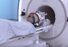
Using 7 Tesla MR techniques pioneered at the University of Minnesota's Center for Magnetic Resonance Research, new insights are being gleaned at sub-millimeter spacial resolutions regarding the actual activities of the human brain.
Now, researchers from the University of Glasgow have taken those techniques and applied them to the complex interaction between what our eyes see — and how the brain perceives it.
The visual cortex is the part of the brain that takes in information from the eyes. That information is conceptualized and contextualized in order for the brain to make sense of what the eyes are looking at.


Ad Statistics
Times Displayed: 45623
Times Visited: 1366 MIT labs, experts in Multi-Vendor component level repair of: MRI Coils, RF amplifiers, Gradient Amplifiers Contrast Media Injectors. System repairs, sub-assembly repairs, component level repairs, refurbish/calibrate. info@mitlabsusa.com/+1 (305) 470-8013
That may sound straightforward enough, but the actual process behind that phenomenon has remained shrouded in mystery.
Previous research into the the nature of this interaction has been limited by an inability to observe the contextual feedback of the visual cortex in an isolated layer of the system, (versus all six cortical layers at once). The researchers from Glasgow said their breakthrough hinges on the fact that input from the retina is mapped out in the visual cortex.
The location from which light hits the eye has a corresponding region in the visual cortex where it is represented, a phenomenon the researchers liken to when light hits a specific portion of a camera's sensor to form a pixel.
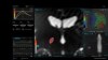
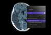
In their study, the researchers showed subjects images, such as that of an automobile, but blocked out a portion of the image with a white square. In the high-definition high-resolution MR image, the absent part of the car was represented in a quieted-down region of voxels in the MR representation.
In this fashion the researchers monitored and mapped out "feedback" and "feedforward" communications between the six layers of the visual cortex, as the brain tried to infer what should have been in the place of the white square.
"Understanding the brain's feedback system is important if we are to develop more powerful computers and artificial intelligence systems, but it might also help us to better understand mental illnesses such as schizophrenia and autism," said Lars Muckli, one of the researchers, and a professor at the University of Glasgow's Institute of Neuroscience & Psychology, in a statement.
"In healthy people, the feedback system plays an important role in understanding concepts and contexts in everyday life. But in people with mental health conditions like autism, we believe that something within the feedback system isn't working correctly. For them everyday scenes that should be regarded as familiar can seem strange," he continued.
Muckli and his colleagues hope that by using MR to get a better understanding of how the visual cortex designs predictive models, they can shed new light on the feeling of unfamiliarity that is typical for many mental health patients. Their research has been published in the journal
Current Biology.