by
Brendon Nafziger, DOTmed News Associate Editor | December 23, 2010

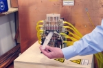
Philips' MPI prototype with
12-cm bore
(Courtesy Philips Research)
Even the most advanced imaging systems, computed tomography and magnetic resonance imaging, have been in use for several decades now. But a new modality that images magnetic particles in the bloodstream and which could help make heart studies more efficient is on the horizon.
Royal Philips Electronics, which invented the technology, said this week it helped launch a German consortium whose goal is to have a whole-body magnetic particle imaging device available for proof-of-concept demonstrations by 2013.
"It's a totally new imaging technology," Steve Klink, a spokesman for Philips, told DOTmed News. "The only thing in common [with MRI] is there are magnets."
The German government has pledged about 10.6 million euros ($13.9 million), about half of the consortium's goal of 20.3 million euros ($26.6 million), which will be paid by the other partners, to develop the demonstration device.
In essence, with magnetic particle imaging, or MPI, the imaging unit detects the magnetic properties of iron oxide nanoparticles - essentially, the same stuff as rust - injected in the body.
It works like this. The nanoparticles are injected into the patient as a bolus, explains Klink, which means the particles don't distribute evenly, but rather travel through the cardiovascular system together as a cloud, perfusing the heart muscle, entering the heart chambers and going through the lungs.
The MPI scanner then detects the magnetic particles, which it quantitatively measures. It can also render a 3-D image, Klink said.
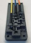
Prototype with 3-cm bore
(Courtesy Philips Research)
Medically, the company is looking to use it either for stress tests or to guide cardiovascular interventions. One of the main possible benefits of the modality is it's sort of an all-in-one system, the company said, doing multiple tests that would normally have to be spread out among different technologies. "With one single measurement, you get information about what's called the ejection fraction, the wall motion, blood flow, speed and also the perfusion of the heart muscles," Klink said.
Decade-old concept
The concept of the technology is almost a decade old, and was dreamt up by MRI researchers Bernhard Gleich and Juergen Weizenecker at Philips' laboratory in Hamburg, Germany. The researchers reasoned that iron-oxide nanoparticles, already used as contrast agents to enhance contrast on MRI images, could be detected outright by a different type of magnetic scanner, possibly boosting the signal-to-noise ratio of the images.