MR-compatible cardiac device implants add significant value to patient care
February 03, 2016
by Lauren Dubinsky, Senior Reporter
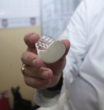
MR-compatible cardiac device implants have been making headlines in recent months as a surge in magnet-friendly pacemakers and implantable cardioverter defibrillators (ICDs) have received regulatory clearance and come to market. The evidence had been established that these devices were safe, but whether or not they were actually useful had yet to be illustrated — until now.
Biotronik received FDA approval for its ProMRI Eluna pacemaker in March of last year. In September Medtronic gained approval for the first MR-compatible ICD, the Evera MRI SureScan ICD System, and in December Biotronik got the nod for administering MR scans to patients with its Iperia ICD system. And that's just to name a few.
First-of-its-kind results have been published for an ongoing study that aims to determine the value of an MR exam for the unique patient population that requires implantable cardiac devices and may also require access to MR exams, (which turns out to be the majority of them). Researchers out of Pittsburgh's Allegheny General Hospital (AGH) have collected the evidence to suggest MR imaging is clearly bringing vital diagnostic value into their case management.
"With the demonstrated safety of MRI for this patient population when performed at cardiac MRI centers with advanced capabilities, and now our research into the effectiveness of using MRI for these patients, it is simply too important a diagnostic tool not to have in our arsenal as we evaluate and determine the best course of care for patients with implanted devices," Dr. Robert Biederman, one of the lead researchers of the study, told HCB News.
MR is an ideal imaging tool because it's a painless test that uses no radiation and allows clinicians to see extremely detailed, 3-D images of organs. Biederman is seeing an increase of almost 6 percent per year in the use of MR for pacemaker patients.
Over the course of several years, the researchers evaluated 157 patients from three implantable cardiac device case groups — 114 neurological, 36 cardiovascular and seven musculoskeletal. They set out to determine whether or not MR imaging added vital insight to diagnosing the various conditions these patients were suffering from.
In 88 percent of the study's neurology cases, the MR scans provided extra value for the final diagnoses and in 18 percent of the cases the MR scan altered the diagnoses entirely. With regard to cardiac and musculoskeletal cases the extra value percentage was even higher; 92 percent and 100 percent, respectively.
In addition, the researchers determined that none of the patients involved in the study experienced any adverse clinical events from undergoing the MR exam.
Out of the more than 200,000 people who have a cardiac pacemaker or cardioverter defibrillator (ICD) implanted each year, about 75 percent will experience a medical situation in their lifetime that could benefit from an MR, according to the University of California, Los Angeles.
There are currently only about 15 to 20 hospitals across the U.S. including AGH that have cardiac MRI centers with the advanced capabilities and experience to pay careful attention to implantable device reprogramming and scanner sequences both before and after the MRI.
At AGH, such patients undergo a comprehensive evaluation of their cardiovascular health and degree of dependence on their device before being imaged. If the patient is cleared, the clinicians perform a baseline device interrogation and put the pacemaker and/or ICD into to a safer mode prior to conducting the exam.
If the patient is found to be non-dependent on the implanted cardiac device under baseline conditions, the device might be turned off completely while the imaging is conducted — which further reduces the risk of device-related complications. The hospital also takes steps to reduce the likelihood of heating, induction of radio-frequency energy, and triggering potentially lethal rhythms.
The patient’s heart rhythms are monitored in real time during the procedures and the entire process is closely overseen by Biederman, a cardiovascular physicist, and a team of nurses and technologists. Once the exams were complete, the implanted devices were reprogrammed to their customary state.
"Most hospitals without cardiac MRI experience must instead use CT ultrasound and catheterization," said Biederman. "These techniques have variable sensitivities and specificities, and they also include radiation exposure, which is not an issue with MRI."
The results of the study were presented last week at the Society of Cardiovascular MRI Scientific Sessions annual meeting in Los Angeles.
