
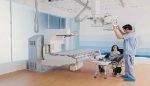
VolumeRad from
GE Healthcare
GE Healthcare
Will tomosynthesis reinvent the X-ray?
November 11, 2015
by Gus Iversen, Editor in Chief
In our modern era of advanced medical imaging capabilities, radiography has settled into having somewhat of a “blue collar” reputation. It is quick, cheap, typically requires no contrast, and is easily accessible — all of which makes X-ray the most utilized medical imaging modality in the world. Denise Taber, radiology manager at Hennepin County Medical Center in Minneapolis, has witnessed 37 years of improvements to radiography, up close and in person. She knows as well as anyone that X-ray is much more than a time-tested exam — it has quietly evolved in its own right while newer and more exotic imaging tools grab the headlines.
By coincidence, Taber spoke to HealthCare Business News on the same day her hospital was cleaning out some old equipment. She and her colleagues shared a laugh watching some old technology going toward the exits (including some old pelvimeters, used to image the birth canal during labor, from the ‘80s) and for a moment were keenly aware of how far they had come. “Maybe 90 percent of our technologists have only learned CR/DR technology, so they don’t remember film and chemicals, hand-processing and all of that,” says Taber. And yet, despite those changes, she says the basic skills and application training for performing an X-ray are basically the same as ever.
As Lori Craig, director of Diagnostic X-ray for Philips, puts it, “We’re still using tube technology to place a shadow image on some type of medium for review.” That hasn’t changed. The transition from film to digital has probably been the biggest single change to radiography — leading to unprecedented speed of acquisition and reductions in dose exposure —but there have been many other changes, too.
For example, in the old days if a clinician needed to X-ray a series of vertebrae, the technicians were required to step in and adjust the system for each individual acquisition. With modern systems, automation and notarization take some of the manual labor out of he examination and free up technicians to do other things.
“In today’s environment where quality scores matter more than ever before, having a tech interface with the patient and making them feel better about the imaging process, that pays dividends to a hospital in a lot of ways,” says Craig, adding that Philips’ DigitalDiagnost radiography rooms were designed with these benefits in mind.
It also means fewer retakes. For aligning the X-ray tube perpendicular to the detector, Pierre Niepel, director of Radiography & Fluoroscopy for Siemens Healthcare, says his company’s MAXalign software automatically displays the angulation to help ensure the equipment and the patient are in the right locations to get the optimal acquisition on the first try.
There are also cost benefits. Craig points out that on average an X-ray costs a hospital roughly $70 whereas an MR exam may be in the range of $1,000 to $1,500. Other imaging exams may be superior for visualizing soft tissue, but for high resolution images of dense structure and bone, or seeing inside the chest cavity, there’s nothing like X-ray. It produces the majority of medical images for just about any hospital, and that’s not likely to change anytime soon.
Fluoroscopy is on the move
Fluoroscopy, a moving image X-ray, is essentially as old as radiography itself. Yet while X-ray has firmly cemented its place as an essential part of medical imaging, fluoroscopy’s success has been somewhat hampered by other advancements. According to Craig, fluoroscopy’s usage fell off in the last five to seven years. “Certain procedures are migrating out to other modalities, be it CT or the bigger interventional cath or vascular labs, where image quality is better because of the technology,” she says. Recently however, it seems to be undergoing a resurgence in popularity. At Hennepin County Medical Center, Taber says that a lull in fluoroscopy exams came to an end roughly three years ago.
“We’re doing different types of exams now, so it’s not so much the barium contrast exams, but it’s more joint injections, pain injections. It seems like we’re doing more arthrograms with MRI follow-up,” she says. For Heidi McIntosh, marketing manager for Global X-ray Solutions at Carestream Health, dynamic flat panel detectors deserve credit for the uptick in fluoroscopy. “Moving from image intensifier to the flat panel detector has made fluoroscopy rooms more valuable because it allows them to be able to do both fluoroscopy and also radiography,” she says.
Her company recently introduced the DRX-Excel and DRX-Excel Plus to meet the needs of clinicians who want the versatility to do both exams with a single system. It’s Carestream’s first foray into fluoroscopy, and she says part of its advantage is that it’s a remote fluoro room instead of a patient-side (or nearby) room. Patient-side fluoroscopy has the image intensifier or detector above the patient and the X-ray source below — but a remote fluoroscopy room flips that around so it’s the same as a r with the image capture happening below, explains McIntosh.
According to Niepel, Siemens is the only OEM that currently offers dynamic flat panel detectors for patient-side fluoroscopy systems. That system, the Luminos Agile Max, can also be customized for radiographic procedures.
According to Michelle Edler, general manager of GE Healthcare’s X-ray Business, speech pathology exams using fluoroscopy are one usage that is on the rise. On the whole, however, her company does not see fluoroscopy experiencing the same kind of momentum they see in radiography. With digital X-ray, going from image intensifiers to flat panels has yielded tremendous workflow benefits, but Edler says she has not seen the same benefits in fluoroscopy. For end users who perform fluoroscopy exams (which could be feeding tube placement, gastrointestinal tract investigations, or other exams), GE manufactures the Precision 500D.
According to Craig, there are multiple moving parts behind the rise in fluoro market activity, one of which is the age of the U.S. install base. “I think it’s a combination of flat panel fluoro which is a technology jump, and the preponderance of old rooms that either cannot dose-track or are just mechanically worn out and need replacement,” she says.
“About 98 percent of the configurations we sell are combo set-up, whether it’s an image intensifier or flat panel room, it’s a combo set up where the customer wants to be able to do radiographic work and fluoro work in the same room, and that allows them to keep their volumes higher and get a better return on investment,” says Craig. Niepel agrees that the staying power of fluoro has a lot to do with the versatility of today’s systems. “A lot of analysts thought it was going to die, but it really didn’t,” he says.
From tomography to tomosynthesis
Tomography was once a fairly common radiography exam. One might have ordered a tomography exam in order to evaluate the lungs, for example, but there were dose concerns associated with it, and eventually better modalities came along and rendered tomography somewhat obsolete. “CT has replaced a lot of that to where we don’t do it, we don’t even have a unit here any longer that can do it,” says Taber at Hennepin. With a CT scan the clinician gets higher image quality, a higher slice count and better dose monitoring. When a CT exam is not possible, tomography can still be justified, says Craig.
“One may argue it was abandoned too early,” says Dr. Bruce Kneeland, section regular X-ray room, chief of the Musculoskeletal Imaging Division and professor of radiology at the Hospital of the University of Pennsylvania. “But it was expensive, difficult to maintain and pretty high radiation.” Kneeland is one of the first practitioners utilizing a new kind of technology that has ascended, in a sense, from tomography and CT. As traditional tomography was a linear process, he is utilizing a new sweeping kind of tomography that moves 30 degrees around the patient, called digital X-ray tomosynthesis.
Unlike tomography, digital X-ray tomosynthesis obtains its acquisition in a single pass and without much more dose exposure than an ordinary radiograph. The value is in being able to see a voluminous image that can be dissected into slices and remove overlap to achieve greater visualization. Many people may think of breast imaging when they hear the word tomosynthesis, but the same imaging principles are being applied to radiography and, with an advanced application called VolumeRAD, GE is among the first OEMs in the emerging market segment. Edler calls X-ray tomosynthesis “the wave of the future.”
Using tomosynthesis, Edler says studies have shown 750x the sensitivity to detecting small-size lung nodules with no loss of specificity. It can also be useful in kidney stone detection. “The resolution you get with X-ray tomosynthesis is at the native level of the detector,” explains Edler. “X-rays typically have higher resolution in the images than CT for example.”
In the orthopedic setting that visualization can be vital for looking at fine anatomical details in wrists or other small bones, in which clinicians want to confirm there is a union building up in the bone. Kneeland’s hospital has been using digital X-ray tomosynthesis for about one year, but for the most part it was only operating in the outpatient orthopedic setting, where its utility was relatively limited. Only in early October did his hospital install one of the systems next to the emergency department.
“It’s more convenient and cheaper than CT, the patient is in the ortho clinic, they come down, they do one scan and we do one pass and you have your study right there,” says Kneeland, adding that the dose exposure is also lower than CT. He and his colleagues are starting a study comparing CT to digital X-ray tomosynthesis. They will look at 300 trauma cases in outpatient settings to compare preliminary diagnosis of the fracture as well as characterizing the fracture. They hope to have 5-10 patients participating per week and some preliminary results by April.
Seeing through walls with dual-energy visualization
The value of X-ray has always been its ability to look noninvasively inside the human body, but sometimes you need to see beyond structures that a normal X-ray cannot clearly see through, such as a bone. Dual-energy imaging makes that possible. “You take two automatic acquisitions, one at low energy and one at high energy,” says Edler. The result is three different perspectives: the traditional X-ray; a bone subtracted image that allows the clinician to focus on soft tissue; and a soft tissue subtracted image to look at the bone.
This can be very useful in trying to identify lung nodules because you don’t have bones obstructing the view of soft tissue. You can also better distinguish calcification. “Non-calcified could indicate a malignant nodule, calcified typically means benign,” says Edler. Philips’ Craig points out that although dual-energy imaging is very useful for certain indications, the challenge is in removing the extra dose from the equation. “Dual-energy by design requires dedicated software, and while it is a small incremental addition of dose to the protocol, it isn’t an incremental dose to the patient because it requires duplicate shots.” Today, some OEMs are meeting this need through post-processing software.
“We [achieve dual-energy imaging] with an algorithm so you only have to take one image,” says Siemens’ Niepel. “We can also do it based on our software-based solution retrospectively.” Besides the dose advantage of this approach, he points to the economy of scale benefit of being able to use the software across an entire range of radiography equipment. For Siemens, having a wide line of radiography tools that are interchangeable and provide the same user experience is fundamental to its business strategy.
Bundled payments may favor radiography
In the U.S., health reform is changing how reimbursement works. Increasing pressure to replace fee-for-procedure billing with bundled payments — in which reimbursement is based on expected costs for clinically-defined episodes of care — will likely impact how physicians order diagnostics.
For Taber, reimbursement has been higher in the previous year than it had been in previous years. “We were nervous there would be a change with chest X-rays this past year and I was nervous we would see a big decrease because that’s a large volume of our work — but we’ve actually been holding fine.” Edler agrees that bundled payments may trigger an uptick in radiography exams, but she also thinks it may soon be time to create a reimbursement code unique to X-ray tomosynthesis. “Right now it fits as an intermediary between X-ray and CT, yet there is no dedicated reimbursement code for that.”
Craig suspects bundled payments may trigger an increase in the amount of fluoro exams being performed. As facilities strive for “fewer touches” per patient, she foresees “a shuffling of the deck of protocols around certain modalities.”
By coincidence, Taber spoke to HealthCare Business News on the same day her hospital was cleaning out some old equipment. She and her colleagues shared a laugh watching some old technology going toward the exits (including some old pelvimeters, used to image the birth canal during labor, from the ‘80s) and for a moment were keenly aware of how far they had come. “Maybe 90 percent of our technologists have only learned CR/DR technology, so they don’t remember film and chemicals, hand-processing and all of that,” says Taber. And yet, despite those changes, she says the basic skills and application training for performing an X-ray are basically the same as ever.
As Lori Craig, director of Diagnostic X-ray for Philips, puts it, “We’re still using tube technology to place a shadow image on some type of medium for review.” That hasn’t changed. The transition from film to digital has probably been the biggest single change to radiography — leading to unprecedented speed of acquisition and reductions in dose exposure —but there have been many other changes, too.
For example, in the old days if a clinician needed to X-ray a series of vertebrae, the technicians were required to step in and adjust the system for each individual acquisition. With modern systems, automation and notarization take some of the manual labor out of he examination and free up technicians to do other things.
“In today’s environment where quality scores matter more than ever before, having a tech interface with the patient and making them feel better about the imaging process, that pays dividends to a hospital in a lot of ways,” says Craig, adding that Philips’ DigitalDiagnost radiography rooms were designed with these benefits in mind.
It also means fewer retakes. For aligning the X-ray tube perpendicular to the detector, Pierre Niepel, director of Radiography & Fluoroscopy for Siemens Healthcare, says his company’s MAXalign software automatically displays the angulation to help ensure the equipment and the patient are in the right locations to get the optimal acquisition on the first try.
There are also cost benefits. Craig points out that on average an X-ray costs a hospital roughly $70 whereas an MR exam may be in the range of $1,000 to $1,500. Other imaging exams may be superior for visualizing soft tissue, but for high resolution images of dense structure and bone, or seeing inside the chest cavity, there’s nothing like X-ray. It produces the majority of medical images for just about any hospital, and that’s not likely to change anytime soon.
Fluoroscopy is on the move
Fluoroscopy, a moving image X-ray, is essentially as old as radiography itself. Yet while X-ray has firmly cemented its place as an essential part of medical imaging, fluoroscopy’s success has been somewhat hampered by other advancements. According to Craig, fluoroscopy’s usage fell off in the last five to seven years. “Certain procedures are migrating out to other modalities, be it CT or the bigger interventional cath or vascular labs, where image quality is better because of the technology,” she says. Recently however, it seems to be undergoing a resurgence in popularity. At Hennepin County Medical Center, Taber says that a lull in fluoroscopy exams came to an end roughly three years ago.
“We’re doing different types of exams now, so it’s not so much the barium contrast exams, but it’s more joint injections, pain injections. It seems like we’re doing more arthrograms with MRI follow-up,” she says. For Heidi McIntosh, marketing manager for Global X-ray Solutions at Carestream Health, dynamic flat panel detectors deserve credit for the uptick in fluoroscopy. “Moving from image intensifier to the flat panel detector has made fluoroscopy rooms more valuable because it allows them to be able to do both fluoroscopy and also radiography,” she says.
Her company recently introduced the DRX-Excel and DRX-Excel Plus to meet the needs of clinicians who want the versatility to do both exams with a single system. It’s Carestream’s first foray into fluoroscopy, and she says part of its advantage is that it’s a remote fluoro room instead of a patient-side (or nearby) room. Patient-side fluoroscopy has the image intensifier or detector above the patient and the X-ray source below — but a remote fluoroscopy room flips that around so it’s the same as a r with the image capture happening below, explains McIntosh.
According to Niepel, Siemens is the only OEM that currently offers dynamic flat panel detectors for patient-side fluoroscopy systems. That system, the Luminos Agile Max, can also be customized for radiographic procedures.
According to Michelle Edler, general manager of GE Healthcare’s X-ray Business, speech pathology exams using fluoroscopy are one usage that is on the rise. On the whole, however, her company does not see fluoroscopy experiencing the same kind of momentum they see in radiography. With digital X-ray, going from image intensifiers to flat panels has yielded tremendous workflow benefits, but Edler says she has not seen the same benefits in fluoroscopy. For end users who perform fluoroscopy exams (which could be feeding tube placement, gastrointestinal tract investigations, or other exams), GE manufactures the Precision 500D.
According to Craig, there are multiple moving parts behind the rise in fluoro market activity, one of which is the age of the U.S. install base. “I think it’s a combination of flat panel fluoro which is a technology jump, and the preponderance of old rooms that either cannot dose-track or are just mechanically worn out and need replacement,” she says.
“About 98 percent of the configurations we sell are combo set-up, whether it’s an image intensifier or flat panel room, it’s a combo set up where the customer wants to be able to do radiographic work and fluoro work in the same room, and that allows them to keep their volumes higher and get a better return on investment,” says Craig. Niepel agrees that the staying power of fluoro has a lot to do with the versatility of today’s systems. “A lot of analysts thought it was going to die, but it really didn’t,” he says.
From tomography to tomosynthesis
Tomography was once a fairly common radiography exam. One might have ordered a tomography exam in order to evaluate the lungs, for example, but there were dose concerns associated with it, and eventually better modalities came along and rendered tomography somewhat obsolete. “CT has replaced a lot of that to where we don’t do it, we don’t even have a unit here any longer that can do it,” says Taber at Hennepin. With a CT scan the clinician gets higher image quality, a higher slice count and better dose monitoring. When a CT exam is not possible, tomography can still be justified, says Craig.
“One may argue it was abandoned too early,” says Dr. Bruce Kneeland, section regular X-ray room, chief of the Musculoskeletal Imaging Division and professor of radiology at the Hospital of the University of Pennsylvania. “But it was expensive, difficult to maintain and pretty high radiation.” Kneeland is one of the first practitioners utilizing a new kind of technology that has ascended, in a sense, from tomography and CT. As traditional tomography was a linear process, he is utilizing a new sweeping kind of tomography that moves 30 degrees around the patient, called digital X-ray tomosynthesis.
Unlike tomography, digital X-ray tomosynthesis obtains its acquisition in a single pass and without much more dose exposure than an ordinary radiograph. The value is in being able to see a voluminous image that can be dissected into slices and remove overlap to achieve greater visualization. Many people may think of breast imaging when they hear the word tomosynthesis, but the same imaging principles are being applied to radiography and, with an advanced application called VolumeRAD, GE is among the first OEMs in the emerging market segment. Edler calls X-ray tomosynthesis “the wave of the future.”
Luminos Agile dual-use
fluoroscopy system
from Siemens Healthcare
fluoroscopy system
from Siemens Healthcare
Using tomosynthesis, Edler says studies have shown 750x the sensitivity to detecting small-size lung nodules with no loss of specificity. It can also be useful in kidney stone detection. “The resolution you get with X-ray tomosynthesis is at the native level of the detector,” explains Edler. “X-rays typically have higher resolution in the images than CT for example.”
In the orthopedic setting that visualization can be vital for looking at fine anatomical details in wrists or other small bones, in which clinicians want to confirm there is a union building up in the bone. Kneeland’s hospital has been using digital X-ray tomosynthesis for about one year, but for the most part it was only operating in the outpatient orthopedic setting, where its utility was relatively limited. Only in early October did his hospital install one of the systems next to the emergency department.
“It’s more convenient and cheaper than CT, the patient is in the ortho clinic, they come down, they do one scan and we do one pass and you have your study right there,” says Kneeland, adding that the dose exposure is also lower than CT. He and his colleagues are starting a study comparing CT to digital X-ray tomosynthesis. They will look at 300 trauma cases in outpatient settings to compare preliminary diagnosis of the fracture as well as characterizing the fracture. They hope to have 5-10 patients participating per week and some preliminary results by April.
Seeing through walls with dual-energy visualization
The value of X-ray has always been its ability to look noninvasively inside the human body, but sometimes you need to see beyond structures that a normal X-ray cannot clearly see through, such as a bone. Dual-energy imaging makes that possible. “You take two automatic acquisitions, one at low energy and one at high energy,” says Edler. The result is three different perspectives: the traditional X-ray; a bone subtracted image that allows the clinician to focus on soft tissue; and a soft tissue subtracted image to look at the bone.
This can be very useful in trying to identify lung nodules because you don’t have bones obstructing the view of soft tissue. You can also better distinguish calcification. “Non-calcified could indicate a malignant nodule, calcified typically means benign,” says Edler. Philips’ Craig points out that although dual-energy imaging is very useful for certain indications, the challenge is in removing the extra dose from the equation. “Dual-energy by design requires dedicated software, and while it is a small incremental addition of dose to the protocol, it isn’t an incremental dose to the patient because it requires duplicate shots.” Today, some OEMs are meeting this need through post-processing software.
“We [achieve dual-energy imaging] with an algorithm so you only have to take one image,” says Siemens’ Niepel. “We can also do it based on our software-based solution retrospectively.” Besides the dose advantage of this approach, he points to the economy of scale benefit of being able to use the software across an entire range of radiography equipment. For Siemens, having a wide line of radiography tools that are interchangeable and provide the same user experience is fundamental to its business strategy.
Bundled payments may favor radiography
In the U.S., health reform is changing how reimbursement works. Increasing pressure to replace fee-for-procedure billing with bundled payments — in which reimbursement is based on expected costs for clinically-defined episodes of care — will likely impact how physicians order diagnostics.
For Taber, reimbursement has been higher in the previous year than it had been in previous years. “We were nervous there would be a change with chest X-rays this past year and I was nervous we would see a big decrease because that’s a large volume of our work — but we’ve actually been holding fine.” Edler agrees that bundled payments may trigger an uptick in radiography exams, but she also thinks it may soon be time to create a reimbursement code unique to X-ray tomosynthesis. “Right now it fits as an intermediary between X-ray and CT, yet there is no dedicated reimbursement code for that.”
Craig suspects bundled payments may trigger an increase in the amount of fluoro exams being performed. As facilities strive for “fewer touches” per patient, she foresees “a shuffling of the deck of protocols around certain modalities.”