
Nanotechnology has already
infiltrated a number of industries,
including medicine.
infiltrated a number of industries,
including medicine.
Cancer Nanomedicine: Tiny Devices Make a Big Difference
October 28, 2009
by Kathy Mahdoubi, Senior Correspondent
This report originally appeared in the October 2009 issue of DOTmed Business News
Imagine devices so miniscule that millions of them can fit into a single cancer cell. This isn't science fiction - it's nanotechnology, and it has already infiltrated a number of industries, including medicine. Several nanoscale pharmaceuticals and biomedical technologies are commercially available, such as antiseptic silver-particle coatings for medical devises and some exciting technologies are still in development, including biosensors that can sense the presence of bacteria, viruses and certain other diseases, but Nanomedicine's biggest research breakthroughs have been in the realm of cancer detection, imaging and treatment. Research institutions across the country and beyond are developing astounding nanotechnologies in the form of cancer biosensors, tumor imaging agents, targeted antitumor drugs and even a minute form of radiation therapy.
The National Science Foundation foresees the nanotechnology industry booming into a $1 trillion a year market by 2015, with nanopharmaceuticals commanding as much as a $180 billion slice of the pie. The term nanomedicine not only indicates the scale of the particles but also the properties of those particles and their particular effect at the cellular, intracellular, molecular and atomic level. By definition, nanoparticles are 1 - 100 nanometers in size. A nanometer is one-billionth of a meter. It helps to think of a nanometer being about 100,000 times thinner than a human hair. Structures that small have some unique advantages when it comes to treating and curing disease.
"Nanomedicine has the potential to create a tremendous paradigm shift that could be described as no less than revolutionary in treating many diseases such as cancer," says Dr. Ahmed Busnaina, director of Nanoscale Science and Engineering Research Center at Northeastern University in Boston, Mass. "If these efforts are successful, it may be possible that no one will die of cancer in the future."
The ultimate cure for cancer is still many years from being realized, but it is definitely obtainable, says Dr. Jie Chen, a researcher and associate professor at the University of Alberta in Edmonton and a principal investigator for the National Institute of Nanotechnology, a Canada-based research and advocacy organization.
"Cancer is not an incurable disease," says Dr. Chen, but both the key and the greatest challenges are early detection and targeted, personalized medicine. "The FDA has approved some nanomedicine already and others are on their way for FDA approval. It's already happening."
Biosensor can smell cancer
Dr. Busnaina's research at Northeastern University is dedicated in part to nanotechnology that can detect certain biological tags indicating disease.
"We focus on micro-biosensors with nanoscale components and structures capable of the simultaneous monitoring of a variety of biomarkers in blood or other biological fluids to assess the progression of disease, toxicity, stress, etc," says Dr. Busnaina.
The university's NSF-funded Nanoscale Science and Engineering Center for High-rate Nanomanufacturing (CHN) is dedicated to developing a process of assembling antibody-coated nanoparticles into "designated nanotrenches" - basically mobile nano pharmacies - that can provide controlled, multi-drug release in real time at the disease site. The research involves the attachment of a tiny chip about 0.1 mm square - smaller than a grain of sand - to a catheter, at which time the nanoparticles are assembled. The process is carried out by "microscopic vision guided micro-manipulators."
"This new nanobio chip design for biomarker monitoring is being tested in vitro and in vivo [using mice and an intravenous catheter] to determine detection limits and effectiveness," says Dr. Busnaina. "The detection limit of our biosensor is almost 1000 times better than current technology."
More nanopharmaceuticals - cancer-drug carriers and delivery systems
This is one of the biggest areas of research and development and several drugs are being "conjugated" with molecules that can protect and add stability to the drug and act as a kind of carrier, while improving blood circulation time and uptake by the body. A variety of particles are being researched to target cancer cells and provide nanoscale therapies.
"Many scientists throughout the world are working in this area," says Dr. Dong M. Shin, a leading investigator for the NIH-funded Center for Cancer Nanotechnology of Excellence and the principal investigator for the Head and Neck Cancer Specialized Program of Research Excellence. Dr. Shin is also a medical oncologist and professor of oncology at Emory University School of Medicine, Atlanta, Ga. and associate director of the university's Winship Cancer Institute.
Some of these kinds of drugs are already on the market or in clinical trials. In fact, several drugs have been approved by the FDA, including Doxil, an intravenous chemotherapy drug that employs liposomes - basically minute fatty bubbles, that help the toxic doxorubicin, an antitumor antibiotic, stay under the radar of white blood cells that might otherwise intervene before the drug can reach the malignant tumor cells. Doxil is prescribed for the treatment of AIDS-related Kaposi's sarcoma, breast cancer and ovarian cancer.
In order to improve cancer cell targeting, researchers are developing cancer drug polymers and attaching ligands - molecules that seek out and bind to the receptors that express a specific gene on the surface of the cancer cell.
"The challenge is determining what kind of receptors we should be targeting," says Dr. Shin. One of the receptors being looked at is EGFR or epidermal growth factor receptor. Although EGFR is over-expressed in cancer cells - which could be used to attract a potential cancer drug - it is also expressed in healthy cells, though it's at a much lower rate than cancer cells. This is not ideal. The ideal is that the appropriate ligand would bind to the cancer cell receptors and enter the cells, at which time enzymes would release the drug and destroy the cancer cell, sparing surrounding healthy tissues because they do not express that specific receptor.
"If we can discover receptors or protein molecules that are exclusively expressed by the cancer cells, those would be our best candidates. But we are still in a state of early development," says Dr. Shin. "The technology is honestly still at the infant stage. We're talking about years or even decades for some of these technologies to come into practical use in the clinic."
Nano-imaging
As powerful as current diagnostic imaging is, oncologists are already behind the curve by the time the slightest tumor can be visualized. A 1 cm tumor mass may contain a proliferation of something in the neighborhood of 10 billion cancer cells, says Dr. Shin. For true early detection, we've got to go smaller.
"If we can detect an aggregate of 100 cells or 1,000 cells with new nanotechnology-based imaging techniques, it would be a huge breakthrough."
Some newer imaging techniques include quantum dot imaging, a kind of molecular imaging that involves fluorescent agents that emit much stronger signals than conventional imaging agents. The signal emitted by these so-called semiconductor crystals is powerful enough to specify that limited number of cancer cells (0.01 mm or less). The dots show up as bright blips on the exam, says Dr. Shin. Certain nanomaterials such as gold or iron oxide can also be used and effectively tracked with MRI or PET scanners.
Nano predators paint the brain and poke holes in tumor cells
Meanwhile, at the University of Washington, researchers are developing cancer-targeting imaging agents using a peptide isolated from the venom of the deathstalker scorpion. The researchers have taken that chlorotoxin and conjugated it with fluorescent nanoparticles that can "paint" tumors in MRI and optical imaging. This agent is specifically engineered for brain cancer tumors and is encapsulated in a biodegradable nanomaterial that allows, for the first time, a nanoimaging agent to pass through the near-impenetrable blood-brain barrier. This agent has the potential to improve current imaging resolutions by a factor of 10 or more, says Dr. Miqin Zhang, professor and nanomedicine researcher at the university.
The nanoimaging agent's cancer targeting ability comes from the chlorotoxin's natural attraction to the expression of matrix metalloproteinase, or MMP2.
"The majority of tumor cells express this MMP2, especially brain tumor cells," says Dr. Zhang. "Chlorotoxin binds to the MMP2 complex. Unlike antibodies that specifically target one kind of cancer, chlorotoxin can see many types of cancer. This is very important, because the brain can have many types of tumors. The chlorotoxin can also inhibit cell invasion, so it has a therapeutic function. Brain tumors are quite invasive and metastasize quickly. If we inject this chlorotoxin we may be able to stop invasion."

The nanoparticle's passage through the blood-brain barrier is made possible by a combination of the particle's diminutive size, chlorotoxin's already proven ability to cross the barrier and with the use of nanomaterials like chitosan, a natural polymer present in crab shells, which helps ease the journey due to its slightly positive charge. Polyethylene glycol makes the whole thing more stable and increases blood circulation time. Typically, molecule-based agents leave the body within minutes, but nanoimaging agents like this one can circulate for hours, says Zhang. The entire nanoparticle is completely biocompatible and biodegradable.
Chlorotoxin is not the only venom-derived toxin being enlisted to disrupt cancer cells. Researchers from Washington University's Siteman Centre of Cancer Nanotechnology Excellence in St. Louis, Mo. have isolated the bee venom toxin melittin, which has been shown to cause tumor cell apoptosis -- cellular suicide.
Dr. Gregory Lanza, professor of medicine and biomedical engineering at Washington University School of Medicine, is a co-investigator on this project.
"Nobody really understands how it works," says Dr. Lanza. "It could be that it induces a better immune response, or it could be antiangiogenesis - it has had some of that effect in some models, or it could be that it's doing something we don't even understand yet."
Angiogenesis is the formation of new blood vessels. Typically there is a frenzy of new vessel formation happening at the growth front of a tumor. Dr. Lanza is principal researcher on another project involving nanodrugs that can halt angiogenesis with the help of a homing ligand isolated from a species of mold called fumagillin. A recent study resulted in an 80% reduction in tumor volume after treatment in rabbits.
Bamboo fungus leads to nano-radiotherapy
At the University of Alberta, Dr. Chen and his colleagues have developed a form of nano-radiotherapy that could one day replace traditional radiation treatments.
"Radiation therapy is like a B-50 bomber - it bombs the whole area. Nano treatment is quite the opposite," says Dr. Chen. "We can specifically target the cells you want to treat. We still use a bomb, but this is like a very small bomb with a GPS system."
A compound naturally occurring in a bamboo-borne fungus gets very excited in the presence of ultrasound. Cancer cells that have taken up nanoparticles conjugated with this compound undergo a kind of cellular radiotherapy.
"This specific bamboo fungus has a resonant frequency that is synchronous with the ultrasound range, which produces free radicals," says Dr. Chen. "The free radicals damage the cells by generating instantaneous radiation - it's basically radiotherapy on a minute scale. The electrons are like satellites and if you shine a certain light or ultrasound on them, it forces them to strike out of their orbit and cause damage to the surrounding DNA."
The bamboo in question grows only in China and Japan and the fungal compound may have originally been used for cosmetic hair removal. Eventually, the compound was found to have unique cancer-fighting properties and was applied in an ointment to suppress skin cancer. Researchers have now taken this compound and conjugated it with nanomaterials that make it water soluble and injectable and have attached either homing ligands that listen for cancer cells' internal clocks, called telomeres, which tick-tock toward infinity in malignant cells, but turn off in healthy cells, or they can attach glucose which is taken up to a greater degree by cancer cells due to their hyper-metabolism.
This nanodrug is currently in human trials for the treatment of prostate cancer, but Dr. Chen says it may come to be used to treat a number of cancers. Radiotherapy at the cellular level still produces side effects similar to traditional radiotherapy, like nausea and vomiting, for instance, but these side effects are greatly diminished and toxicity is low.
Driving nanomedicine forward
Chen was in attendance at the 2009 NanoWeek held in April and hosted by the National Institutes of Health at their Bethesda, Md. campus. "The NIH has listed nanomedicine as one of their strategic roadmaps for the next decade," says Chen. "They are very keen on the development of nanomedicine."
The NIH has provided a significant amount of funding toward nanotechnology, including nanomedicine and eight research centers for the development of nanomedicine have been set up across the country. The NIH's strategic roadmap sets the bar for innovations to come.
"The NIH Roadmap Nanomedicine Initiative is one of several initiatives within the NIH Roadmap that is funded by the common fund - not any one institute," explains Richard S. Fisher, Director of Program Planning and Analysis for the initiative. "This represents a small portion of the total NIH investment in nanotechnology."
To learn more about the newest developments in nanomedicine, check out the American Society of Nanomedicine's inaugural conference scheduled to be held October 22-25 at Bolger Center in Potomac, Md.
Imagine devices so miniscule that millions of them can fit into a single cancer cell. This isn't science fiction - it's nanotechnology, and it has already infiltrated a number of industries, including medicine. Several nanoscale pharmaceuticals and biomedical technologies are commercially available, such as antiseptic silver-particle coatings for medical devises and some exciting technologies are still in development, including biosensors that can sense the presence of bacteria, viruses and certain other diseases, but Nanomedicine's biggest research breakthroughs have been in the realm of cancer detection, imaging and treatment. Research institutions across the country and beyond are developing astounding nanotechnologies in the form of cancer biosensors, tumor imaging agents, targeted antitumor drugs and even a minute form of radiation therapy.
The National Science Foundation foresees the nanotechnology industry booming into a $1 trillion a year market by 2015, with nanopharmaceuticals commanding as much as a $180 billion slice of the pie. The term nanomedicine not only indicates the scale of the particles but also the properties of those particles and their particular effect at the cellular, intracellular, molecular and atomic level. By definition, nanoparticles are 1 - 100 nanometers in size. A nanometer is one-billionth of a meter. It helps to think of a nanometer being about 100,000 times thinner than a human hair. Structures that small have some unique advantages when it comes to treating and curing disease.
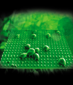
Carbon nanofibers serve as
gene delivery tools
(Image courtesy of Tim McKnight
at the Oak Ridge National
Laboratory)
gene delivery tools
(Image courtesy of Tim McKnight
at the Oak Ridge National
Laboratory)
"Nanomedicine has the potential to create a tremendous paradigm shift that could be described as no less than revolutionary in treating many diseases such as cancer," says Dr. Ahmed Busnaina, director of Nanoscale Science and Engineering Research Center at Northeastern University in Boston, Mass. "If these efforts are successful, it may be possible that no one will die of cancer in the future."
The ultimate cure for cancer is still many years from being realized, but it is definitely obtainable, says Dr. Jie Chen, a researcher and associate professor at the University of Alberta in Edmonton and a principal investigator for the National Institute of Nanotechnology, a Canada-based research and advocacy organization.
"Cancer is not an incurable disease," says Dr. Chen, but both the key and the greatest challenges are early detection and targeted, personalized medicine. "The FDA has approved some nanomedicine already and others are on their way for FDA approval. It's already happening."
Biosensor can smell cancer
Dr. Busnaina's research at Northeastern University is dedicated in part to nanotechnology that can detect certain biological tags indicating disease.
"We focus on micro-biosensors with nanoscale components and structures capable of the simultaneous monitoring of a variety of biomarkers in blood or other biological fluids to assess the progression of disease, toxicity, stress, etc," says Dr. Busnaina.
The university's NSF-funded Nanoscale Science and Engineering Center for High-rate Nanomanufacturing (CHN) is dedicated to developing a process of assembling antibody-coated nanoparticles into "designated nanotrenches" - basically mobile nano pharmacies - that can provide controlled, multi-drug release in real time at the disease site. The research involves the attachment of a tiny chip about 0.1 mm square - smaller than a grain of sand - to a catheter, at which time the nanoparticles are assembled. The process is carried out by "microscopic vision guided micro-manipulators."
"This new nanobio chip design for biomarker monitoring is being tested in vitro and in vivo [using mice and an intravenous catheter] to determine detection limits and effectiveness," says Dr. Busnaina. "The detection limit of our biosensor is almost 1000 times better than current technology."
More nanopharmaceuticals - cancer-drug carriers and delivery systems
This is one of the biggest areas of research and development and several drugs are being "conjugated" with molecules that can protect and add stability to the drug and act as a kind of carrier, while improving blood circulation time and uptake by the body. A variety of particles are being researched to target cancer cells and provide nanoscale therapies.
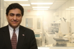
Dr. Ahmed Busnaina, director of
the Nanoscale Science and
Engineering Research Center at
Northeastern University in Boston,
at a lab at the Center for
High-rate Nanomanufacturing
the Nanoscale Science and
Engineering Research Center at
Northeastern University in Boston,
at a lab at the Center for
High-rate Nanomanufacturing
"Many scientists throughout the world are working in this area," says Dr. Dong M. Shin, a leading investigator for the NIH-funded Center for Cancer Nanotechnology of Excellence and the principal investigator for the Head and Neck Cancer Specialized Program of Research Excellence. Dr. Shin is also a medical oncologist and professor of oncology at Emory University School of Medicine, Atlanta, Ga. and associate director of the university's Winship Cancer Institute.
Some of these kinds of drugs are already on the market or in clinical trials. In fact, several drugs have been approved by the FDA, including Doxil, an intravenous chemotherapy drug that employs liposomes - basically minute fatty bubbles, that help the toxic doxorubicin, an antitumor antibiotic, stay under the radar of white blood cells that might otherwise intervene before the drug can reach the malignant tumor cells. Doxil is prescribed for the treatment of AIDS-related Kaposi's sarcoma, breast cancer and ovarian cancer.
In order to improve cancer cell targeting, researchers are developing cancer drug polymers and attaching ligands - molecules that seek out and bind to the receptors that express a specific gene on the surface of the cancer cell.
"The challenge is determining what kind of receptors we should be targeting," says Dr. Shin. One of the receptors being looked at is EGFR or epidermal growth factor receptor. Although EGFR is over-expressed in cancer cells - which could be used to attract a potential cancer drug - it is also expressed in healthy cells, though it's at a much lower rate than cancer cells. This is not ideal. The ideal is that the appropriate ligand would bind to the cancer cell receptors and enter the cells, at which time enzymes would release the drug and destroy the cancer cell, sparing surrounding healthy tissues because they do not express that specific receptor.
"If we can discover receptors or protein molecules that are exclusively expressed by the cancer cells, those would be our best candidates. But we are still in a state of early development," says Dr. Shin. "The technology is honestly still at the infant stage. We're talking about years or even decades for some of these technologies to come into practical use in the clinic."
Nano-imaging
As powerful as current diagnostic imaging is, oncologists are already behind the curve by the time the slightest tumor can be visualized. A 1 cm tumor mass may contain a proliferation of something in the neighborhood of 10 billion cancer cells, says Dr. Shin. For true early detection, we've got to go smaller.
"If we can detect an aggregate of 100 cells or 1,000 cells with new nanotechnology-based imaging techniques, it would be a huge breakthrough."
Some newer imaging techniques include quantum dot imaging, a kind of molecular imaging that involves fluorescent agents that emit much stronger signals than conventional imaging agents. The signal emitted by these so-called semiconductor crystals is powerful enough to specify that limited number of cancer cells (0.01 mm or less). The dots show up as bright blips on the exam, says Dr. Shin. Certain nanomaterials such as gold or iron oxide can also be used and effectively tracked with MRI or PET scanners.
Nano predators paint the brain and poke holes in tumor cells
Meanwhile, at the University of Washington, researchers are developing cancer-targeting imaging agents using a peptide isolated from the venom of the deathstalker scorpion. The researchers have taken that chlorotoxin and conjugated it with fluorescent nanoparticles that can "paint" tumors in MRI and optical imaging. This agent is specifically engineered for brain cancer tumors and is encapsulated in a biodegradable nanomaterial that allows, for the first time, a nanoimaging agent to pass through the near-impenetrable blood-brain barrier. This agent has the potential to improve current imaging resolutions by a factor of 10 or more, says Dr. Miqin Zhang, professor and nanomedicine researcher at the university.
The nanoimaging agent's cancer targeting ability comes from the chlorotoxin's natural attraction to the expression of matrix metalloproteinase, or MMP2.
"The majority of tumor cells express this MMP2, especially brain tumor cells," says Dr. Zhang. "Chlorotoxin binds to the MMP2 complex. Unlike antibodies that specifically target one kind of cancer, chlorotoxin can see many types of cancer. This is very important, because the brain can have many types of tumors. The chlorotoxin can also inhibit cell invasion, so it has a therapeutic function. Brain tumors are quite invasive and metastasize quickly. If we inject this chlorotoxin we may be able to stop invasion."
High-resolution transmission
electron microscopy of bamboo
fungus coated with nanomaterials
(Image courtesy of the
University of Alberta)
electron microscopy of bamboo
fungus coated with nanomaterials
(Image courtesy of the
University of Alberta)
The nanoparticle's passage through the blood-brain barrier is made possible by a combination of the particle's diminutive size, chlorotoxin's already proven ability to cross the barrier and with the use of nanomaterials like chitosan, a natural polymer present in crab shells, which helps ease the journey due to its slightly positive charge. Polyethylene glycol makes the whole thing more stable and increases blood circulation time. Typically, molecule-based agents leave the body within minutes, but nanoimaging agents like this one can circulate for hours, says Zhang. The entire nanoparticle is completely biocompatible and biodegradable.
Chlorotoxin is not the only venom-derived toxin being enlisted to disrupt cancer cells. Researchers from Washington University's Siteman Centre of Cancer Nanotechnology Excellence in St. Louis, Mo. have isolated the bee venom toxin melittin, which has been shown to cause tumor cell apoptosis -- cellular suicide.
Dr. Gregory Lanza, professor of medicine and biomedical engineering at Washington University School of Medicine, is a co-investigator on this project.
"Nobody really understands how it works," says Dr. Lanza. "It could be that it induces a better immune response, or it could be antiangiogenesis - it has had some of that effect in some models, or it could be that it's doing something we don't even understand yet."
Angiogenesis is the formation of new blood vessels. Typically there is a frenzy of new vessel formation happening at the growth front of a tumor. Dr. Lanza is principal researcher on another project involving nanodrugs that can halt angiogenesis with the help of a homing ligand isolated from a species of mold called fumagillin. A recent study resulted in an 80% reduction in tumor volume after treatment in rabbits.
Bamboo fungus leads to nano-radiotherapy
At the University of Alberta, Dr. Chen and his colleagues have developed a form of nano-radiotherapy that could one day replace traditional radiation treatments.
"Radiation therapy is like a B-50 bomber - it bombs the whole area. Nano treatment is quite the opposite," says Dr. Chen. "We can specifically target the cells you want to treat. We still use a bomb, but this is like a very small bomb with a GPS system."
A compound naturally occurring in a bamboo-borne fungus gets very excited in the presence of ultrasound. Cancer cells that have taken up nanoparticles conjugated with this compound undergo a kind of cellular radiotherapy.
"This specific bamboo fungus has a resonant frequency that is synchronous with the ultrasound range, which produces free radicals," says Dr. Chen. "The free radicals damage the cells by generating instantaneous radiation - it's basically radiotherapy on a minute scale. The electrons are like satellites and if you shine a certain light or ultrasound on them, it forces them to strike out of their orbit and cause damage to the surrounding DNA."
The bamboo in question grows only in China and Japan and the fungal compound may have originally been used for cosmetic hair removal. Eventually, the compound was found to have unique cancer-fighting properties and was applied in an ointment to suppress skin cancer. Researchers have now taken this compound and conjugated it with nanomaterials that make it water soluble and injectable and have attached either homing ligands that listen for cancer cells' internal clocks, called telomeres, which tick-tock toward infinity in malignant cells, but turn off in healthy cells, or they can attach glucose which is taken up to a greater degree by cancer cells due to their hyper-metabolism.
This nanodrug is currently in human trials for the treatment of prostate cancer, but Dr. Chen says it may come to be used to treat a number of cancers. Radiotherapy at the cellular level still produces side effects similar to traditional radiotherapy, like nausea and vomiting, for instance, but these side effects are greatly diminished and toxicity is low.
Driving nanomedicine forward
Chen was in attendance at the 2009 NanoWeek held in April and hosted by the National Institutes of Health at their Bethesda, Md. campus. "The NIH has listed nanomedicine as one of their strategic roadmaps for the next decade," says Chen. "They are very keen on the development of nanomedicine."
The NIH has provided a significant amount of funding toward nanotechnology, including nanomedicine and eight research centers for the development of nanomedicine have been set up across the country. The NIH's strategic roadmap sets the bar for innovations to come.
"The NIH Roadmap Nanomedicine Initiative is one of several initiatives within the NIH Roadmap that is funded by the common fund - not any one institute," explains Richard S. Fisher, Director of Program Planning and Analysis for the initiative. "This represents a small portion of the total NIH investment in nanotechnology."
To learn more about the newest developments in nanomedicine, check out the American Society of Nanomedicine's inaugural conference scheduled to be held October 22-25 at Bolger Center in Potomac, Md.